Toward Better Antibiotics: Crystallographic Studies of a Novel Class of DD-Peptidase/beta-Lactamase Inhibitors.
Silvaggi, N.R., Kaur, K., Adediran, S.A., Pratt, R.F., Kelly, J.A.(2004) Biochemistry 43: 7046-7053
- PubMed: 15170342 Search on PubMed
- DOI: https://doi.org/10.1021/bi049612c
- Primary Citation Related Structures:
1SCW, 1SDE - PubMed Abstract:
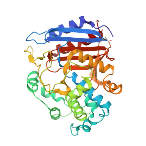
Beta-lactam antibiotics are vital weapons in the treatment of bacterial infections, but their future is under increasing threat from beta-lactamases. These bacterial enzymes hydrolyze and inactivate beta-lactam antibiotics, rendering the host cell resistant to the bactericidal effects of the drugs. Nevertheless, the bacterial D-alanyl-D-alanine transpeptidases (DD-peptidases), the killing targets of beta-lactams, remain attractive targets for antibiotic compounds. Cyclic acyl phosph(on)ates have been developed and investigated as potential inhibitors of both transpeptidases and beta-lactamases. The X-ray crystal structures of the complexes of the Streptomyces strain R61 DD-peptidase inhibited by a bicyclic [1-hydroxy-4,5-benzo-2,6-dioxaphosphorinanone(3)-1-oxide] and a monocyclic [1-hydroxy-4-phenyl-2,6-dioxaphosphorinanone(3)-1-oxide] acyl phosphate were determined to investigate the mode of action of these novel inhibitors. The structures show, first, that these inhibitors form covalent bonds with the active site serine residue of the enzyme and that the refractory complexes thus formed are phosphoryl-enzyme species rather than acyl enzymes. The complexes are long-lived largely because, after ring opening, the ligands adopt conformations that cannot directly recyclize, the latter a phenomenon previously observed with cyclic acyl phosph(on)ates. While the two inhibitors bind in nearly identical conformations, the phosphoryl-enzyme complex formed from the monocyclic compound is significantly less mobile than that formed from the bicyclic compound. Despite this difference, the complex with the bicyclic compound breaks down to regenerate free enzyme somewhat more slowly than that of the monocyclic. This may be because of steric problems associated with the reorientation of the larger bicyclic ligand required for reactivation. The structures are strikingly different in the orientation of the phosphoryl moiety from those generated using more specific phosph(on)ates. Models of the noncovalent complexes of the monocyclic compound with the R61 DD-peptidase and a structurally very similar class C beta-lactamase suggest reasons why the former enzyme is phosphorylated by this compound, while the latter is acylated. Finally, this paper provides information that will help in the design of additional DD-peptidase inhibitors with the potential to serve as leads in the development of novel antibiotics.
- Department of Molecular and Cell Biology and Institute for Materials Science, University of Connecticut, Storrs, Connecticut 06269-3125, USA.
Organizational Affiliation: