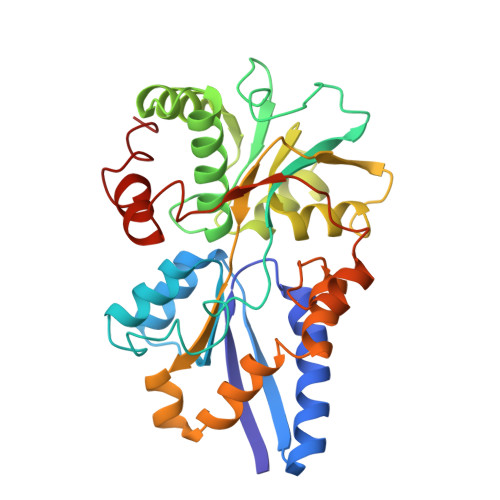
1SBP

1.7 ANGSTROMS REFINED STRUCTURE OF SULFATE-BINDING PROTEIN INVOLVED IN ACTIVE TRANSPORT AND NOVEL MODE OF SULFATE BINDING


Experimental Data Snapshot

  • Method: X-RAY DIFFRACTION
  • Resolution: 1.70 Å
  • R-Value Observed: 0.178 

wwPDB Validation   3D Report Full Report


This is version 1.3 of the entry. See complete history


Literature

Dominant role of local dipoles in stabilizing uncompensated charges on a sulfate sequestered in a periplasmic active transport protein.

He, J.J.Quiocho, F.A.

(1993) Protein Sci 2: 1643-1647

  • DOI: https://doi.org/10.1002/pro.5560021010
  • Primary Citation of Related Structures:  
    1SBP

  • PubMed Abstract: 

    Electrostatic interactions are among the key factors determining the structure and function of proteins. Here we report experimental results that illuminate the functional importance of local dipoles to these interactions. The refined 1.7-A X-ray structure of the liganded form of the sulfate-binding protein, a primary sulfate active transport receptor of Salmonella typhimurium, shows that the sulfate dianion is completely buried and bound by hydrogen bonds (mostly main-chain peptide NH groups) and van der Waals forces. The sulfate is also closely linked, via one of these peptide units, to a His residue. It is also adjacent to the N-termini of three alpha-helices, of which the two shortest have their C-termini "capped" by Arg residues. Site-directed mutagenesis of the recombinant Escherichia coli sulfate receptor had no effect on sulfate-binding activity when an Asn residue was substituted for the positively charged His and the two Arg (changed singly and together) residues. These results, combined with other observations, further solidify the idea that stabilization of uncompensated charges in a protein is a highly localized process that involves a collection of local dipoles, including those of peptide units confined to the first turns of helices. The contribution of helix macrodipoles appears insignificant.


  • Organizational Affiliation

    Howard Hughes Medical Institute, Baylor College of Medicine, Houston, Texas 77030.


Macromolecules
Find similar proteins by:  (by identity cutoff)  |  3D Structure
Entity ID: 1
MoleculeChains Sequence LengthOrganismDetailsImage
SULFATE-BINDING PROTEIN310Salmonella enterica subsp. enterica serovar TyphimuriumMutation(s): 0 
UniProt
Find proteins for P02906 (Salmonella typhimurium (strain LT2 / SGSC1412 / ATCC 700720))
Explore P02906 
Go to UniProtKB:  P02906
Entity Groups  
Sequence Clusters30% Identity50% Identity70% Identity90% Identity95% Identity100% Identity
UniProt GroupP02906
Sequence Annotations
Expand
  • Reference Sequence
Small Molecules
Ligands 1 Unique
IDChains Name / Formula / InChI Key2D Diagram3D Interactions
SO4
Query on SO4

Download Ideal Coordinates CCD File 
B [auth A]SULFATE ION
O4 S
QAOWNCQODCNURD-UHFFFAOYSA-L
Experimental Data & Validation

Experimental Data

  • Method: X-RAY DIFFRACTION
  • Resolution: 1.70 Å
  • R-Value Observed: 0.178 
  • Space Group: P 21 21 21
Unit Cell:
Length ( Å )Angle ( ˚ )
a = 40.77α = 90
b = 49.37β = 90
c = 141.03γ = 90
Software Package:
Software NamePurpose
PROLSQrefinement

Structure Validation

View Full Validation Report


Entry History 

Deposition Data

Revision History  (Full details and data files)

  • Version 1.0: 1993-10-31
    Type: Initial release
  • Version 1.1: 2008-03-24
    Changes: Version format compliance
  • Version 1.2: 2011-07-13
    Changes: Version format compliance
  • Version 1.3: 2024-02-14
    Changes: Data collection, Database references, Derived calculations, Other