1SBN | pdb_00001sbn

REFINED CRYSTAL STRUCTURES OF SUBTILISIN NOVO IN COMPLEX WITH WILD-TYPE AND TWO MUTANT EGLINS. COMPARISON WITH OTHER SERINE PROTEINASE INHIBITOR COMPLEXES


Experimental Data Snapshot

  • Method: X-RAY DIFFRACTION
  • Resolution: 2.10 Å
  • R-Value Observed: 
    0.186 (Depositor) 

wwPDB Validation 3D Report Full Report

Validation slider image for 1SBN

This is version 1.4 of the entry. See complete history

Literature

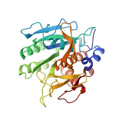
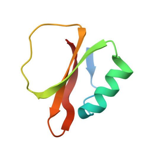
Refined crystal structures of subtilisin novo in complex with wild-type and two mutant eglins. Comparison with other serine proteinase inhibitor complexes.

Heinz, D.W.Priestle, J.P.Rahuel, J.Wilson, K.S.Grutter, M.G.

(1991) J Mol Biology 217: 353-371

  • DOI: https://doi.org/10.1016/0022-2836(91)90549-l
  • Primary Citation Related Structures: 
    1SBN, 1SIB

  • PubMed Abstract: 

    The crystal structures of the complexes formed between subtilisin Novo and three inhibitors, eglin c, Arg45-eglin c and Lys53-eglin c have been determined using molecular replacement and difference Fourier techniques and refined at 2.4 A, 2.1 A, and 2.4 A resolution, respectively. The mutants Arg45-eglin c and Lys53-eglin c were constructed by site-directed mutagenesis in order to investigate the inhibitory specificity and stability of eglin c. Arg45-eglin became a potent trypsin inhibitor, in contrast to native eglin, which is an elastase inhibitor. This specificity change was rationalized by comparing the structures of Arg45-eglin and basic pancreatic trypsin inhibitor and their interactions with trypsin. The residue Arg53, which participates in a complex network of hydrogen bonds formed between the core and the binding loop of eglin c, was replaced with the shorter basic amino acid lysine in the mutant Lys53-eglin. Two hydrogen bonds with Thr44, located in the binding loop, can no longer be formed but are partially restored by a water molecule bound in the vicinity of Lys53. Eglin c in complexes with both subtilisin Novo and subtilisin Carlsberg was crystallized in two different space groups. Comparison of the complexes showed a rigid body rotation for the eglin c core of 11.5 degrees with respect to the enzyme, probably caused by different intermolecular contacts in both crystal forms.


  • Organizational Affiliation
    • Pharmaceutical Division, Ciba-Geigy Ltd., Basel, Switzerland.

Macromolecule Content 

  • Total Structure Weight: 35.78 kDa 
  • Atom Count: 2,781 
  • Modeled Residue Count: 338 
  • Deposited Residue Count: 345 
  • Unique protein chains: 2

Macromolecules

Find similar proteins by:|  3D Structure
Entity ID: 1
MoleculeChains  Sequence LengthOrganismDetailsImage
SUBTILISIN NOVO BPN'A [auth E]275Bacillus subtilisMutation(s): 0 
EC: 3.4.21.62
UniProt
Find proteins for P00782 (Bacillus amyloliquefaciens)
Explore P00782 
Go to UniProtKB:  P00782
Entity Groups
Sequence Clusters30% Identity50% Identity70% Identity90% Identity95% Identity100% Identity
UniProt GroupP00782
Sequence Annotations
Expand
Reference Sequence
Find similar proteins by:|  3D Structure
Entity ID: 2
MoleculeChains  Sequence LengthOrganismDetailsImage
EGLIN CB [auth I]70Hirudo medicinalisMutation(s): 0 
UniProt
Find proteins for P01051 (Hirudo medicinalis)
Explore P01051 
Go to UniProtKB:  P01051
Entity Groups
Sequence Clusters30% Identity50% Identity70% Identity90% Identity95% Identity100% Identity
UniProt GroupP01051
Sequence Annotations
Expand
Reference Sequence

Small Molecules

Experimental Data & Validation

Experimental Data

  • Method: X-RAY DIFFRACTION
  • Resolution: 2.10 Å
  • R-Value Observed: 0.186 (Depositor) 
Space Group: P 31 2 1
Unit Cell:
Length ( Å )Angle ( ˚ )
a = 84.9α = 90
b = 84.9β = 90
c = 89.1γ = 120
Software Package:
Software NamePurpose
PROLSQrefinement

Structure Validation

View Full Validation Report


Entry History 

Deposition Data

Revision History  (Full details and data files)

  • Version 1.0: 1994-01-31
    Type: Initial release
  • Version 1.1: 2008-03-03
    Changes: Version format compliance
  • Version 1.2: 2011-07-13
    Changes: Version format compliance
  • Version 1.3: 2017-11-29
    Changes: Derived calculations, Other
  • Version 1.4: 2024-02-14
    Changes: Data collection, Database references, Derived calculations