1SAZ | pdb_00001saz

Membership in the ASKHA Superfamily: Enzymological Properties and Crystal Structure of Butyrate Kinase 2 from Thermotoga maritima


Experimental Data Snapshot

  • Method: X-RAY DIFFRACTION
  • Resolution: 2.50 Å
  • R-Value Free: 
    0.267 (Depositor), 0.266 (DCC) 
  • R-Value Work: 
    0.217 (Depositor), 0.216 (DCC) 

wwPDB Validation 3D Report Full Report

Validation slider image for 1SAZ

Ligand Structure Quality Assessment 


This is version 1.3 of the entry. See complete history

Literature

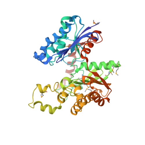
Crystal structure of butyrate kinase 2 from Thermotoga maritima, a member of the ASKHA superfamily of phosphotransferases.

Diao, J.Hasson, M.S.

(2009) J Bacteriol 191: 2521-2529

  • DOI: https://doi.org/10.1128/JB.00906-08
  • Primary Citation Related Structures: 
    1SAZ

  • PubMed Abstract: 

    The enzymatic transfer of phosphoryl groups is central to the control of many cellular processes. One of the phosphoryl transfer mechanisms, that of acetate kinase, is not completely understood. Besides better understanding of the mechanism of acetate kinase, knowledge of the structure of butyrate kinase 2 (Buk2) will aid in the interpretation of active-site structure and provide information on the structural basis of substrate specificity. The gene buk2 from Thermotoga maritima encodes a member of the ASKHA (acetate and sugar kinases/heat shock cognate/actin) superfamily of phosphotransferases. The encoded protein Buk2 catalyzes the phosphorylation of butyrate and isobutyrate. We have determined the 2.5-A crystal structure of Buk2 complexed with (beta,gamma-methylene) adenosine 5'-triphosphate. Buk2 folds like an open-shelled clam, with each of the two domains representing one of the two shells. In the open active-site cleft between the N- and C-terminal domains, the active-site residues consist of two histidines, two arginines, and a cluster of hydrophobic residues. The ATP binding region of Buk2 in the C-terminal domain consists of abundant glycines for nucleotide binding, and the ATP binding motif is similar to those of other members of the ASKHA superfamily. The enzyme exists as an octamer, in which four disulfide bonds form between intermolecular cysteines. Sequence alignment and structure superposition identify the simplicity of the monomeric Buk2 structure, a probable substrate binding site, the key residues in catalyzing phosphoryl transfer, and the substrate specificity differences among Buk2, acetate, and propionate kinases. The possible enzyme mechanisms are discussed.


  • Organizational Affiliation
    • Department of Biological Sciences, Purdue University, West Lafayette, Indiana 47907, USA. diaojs@netscape.net

Macromolecule Content 

  • Total Structure Weight: 44.19 kDa 
  • Atom Count: 3,065 
  • Modeled Residue Count: 375 
  • Deposited Residue Count: 381 
  • Unique protein chains: 1

Macromolecules

Find similar proteins by:|  3D Structure
Entity ID: 1
MoleculeChains  Sequence LengthOrganismDetailsImage
Probable butyrate kinase 2381Thermotoga maritimaMutation(s): 12 
Gene Names: BUK2TM1756
EC: 2.7.2.7
UniProt
Find proteins for Q9X278 (Thermotoga maritima (strain ATCC 43589 / DSM 3109 / JCM 10099 / NBRC 100826 / MSB8))
Explore Q9X278 
Go to UniProtKB:  Q9X278
Entity Groups
Sequence Clusters30% Identity50% Identity70% Identity90% Identity95% Identity100% Identity
UniProt GroupQ9X278
Sequence Annotations
Expand
Reference Sequence

Small Molecules

Modified Residues  1 Unique
IDChains TypeFormula2D DiagramParent
MSE
Query on MSE
A
L-PEPTIDE LINKINGC5 H11 N O2 SeMET

Experimental Data & Validation

Experimental Data

  • Method: X-RAY DIFFRACTION
  • Resolution: 2.50 Å
  • R-Value Free:  0.267 (Depositor), 0.266 (DCC) 
  • R-Value Work:  0.217 (Depositor), 0.216 (DCC) 
Space Group: I 4 2 2
Unit Cell:
Length ( Å )Angle ( ˚ )
a = 197.685α = 90
b = 197.685β = 90
c = 58.238γ = 90
Software Package:
Software NamePurpose
DENZOdata reduction
SCALEPACKdata scaling
SOLVEphasing
CNSrefinement

Structure Validation

View Full Validation Report


Ligand Structure Quality Assessment 


Entry History 

Deposition Data

Revision History  (Full details and data files)

  • Version 1.0: 2005-03-29
    Type: Initial release
  • Version 1.1: 2008-04-29
    Changes: Version format compliance
  • Version 1.2: 2011-07-13
    Changes: Version format compliance
  • Version 1.3: 2024-11-13
    Changes: Data collection, Database references, Derived calculations, Structure summary