1S6L | pdb_00001s6l

Solution structure of MerB, the Organomercurial Lyase involved in the bacterial mercury resistance system


Experimental Data Snapshot

  • Method: SOLUTION NMR
  • Conformers Calculated: 55 
  • Conformers Submitted: 20 
  • Selection Criteria: The submitted models are the 20 structures with no upper bound violation greater that 0.3 armstrongs and no dihedral angle restraint violation greater than 2 degrees and with the lowest energies. 

wwPDB Validation 3D Report Full Report

Validation slider image for 1S6L

This is version 1.3 of the entry. See complete history

Literature

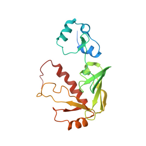
NMR structural studies reveal a novel protein fold for MerB, the organomercurial lyase involved in the bacterial mercury resistance system.

Di Lello, P.Benison, G.C.Valafar, H.Pitts, K.E.Summers, A.O.Legault, P.Omichinski, J.G.

(2004) Biochemistry 43: 8322-8332

  • DOI: https://doi.org/10.1021/bi049669z
  • Primary Citation Related Structures: 
    1S6L

  • PubMed Abstract: 

    Mercury resistant bacteria have developed a system of two enzymes (MerA and MerB), which allows them to efficiently detoxify both ionic and organomercurial compounds. The organomercurial lyase (MerB) catalyzes the protonolysis of the carbon-mercury bond resulting in the formation of ionic mercury and a reduced hydrocarbon. The ionic mercury [Hg(II)] is subsequently reduced to the less reactive elemental mercury [Hg(0)] by a specific mercuric reductase (MerA). To better understand MerB's unique enzymatic activity, we used nuclear magnetic resonance (NMR) spectroscopy to determine the structure of the free enzyme. MerB is characterized by a novel protein fold consisting of three noninteracting antiparallel beta-sheets surrounded by six alpha-helices. By comparing the NMR data of free MerB and the MerB/Hg/DTT complex, we identified a set of residues that likely define a Hg/DTT binding site. These residues cluster around two cysteines (C(96) and C(159)) that are crucial to MerB's catalytic activity. A detailed analysis of the structure revealed the presence of an extensive hydrophobic groove adjacent to this Hg/DTT binding site. This extensive hydrophobic groove has the potential to interact with the hydrocarbon moiety of a wide variety of substrates and may explain the broad substrate specificity of MerB.


  • Organizational Affiliation
    • Department of Biochemistry & Molecular Biology, University of Georgia, Athens, Georgia 30602, USA.

Macromolecule Content 

  • Total Structure Weight: 23.06 kDa 
  • Atom Count: 1,466 
  • Modeled Residue Count: 192 
  • Deposited Residue Count: 212 
  • Unique protein chains: 1

Macromolecules

Find similar proteins by:|  3D Structure
Entity ID: 1
MoleculeChains  Sequence LengthOrganismDetailsImage
Alkylmercury lyase212Escherichia coliMutation(s): 0 
Gene Names: MERB
EC: 4.99.1.2
UniProt
Find proteins for P77072 (Escherichia coli)
Explore P77072 
Go to UniProtKB:  P77072
Entity Groups
Sequence Clusters30% Identity50% Identity70% Identity90% Identity95% Identity100% Identity
UniProt GroupP77072
Sequence Annotations
Expand
Reference Sequence

Experimental Data & Validation

Experimental Data

  • Method: SOLUTION NMR
  • Conformers Calculated: 55 
  • Conformers Submitted: 20 
  • Selection Criteria: The submitted models are the 20 structures with no upper bound violation greater that 0.3 armstrongs and no dihedral angle restraint violation greater than 2 degrees and with the lowest energies. 

Structure Validation

View Full Validation Report


Entry History 

Deposition Data

Revision History  (Full details and data files)

  • Version 1.0: 2005-04-19
    Type: Initial release
  • Version 1.1: 2008-04-29
    Changes: Version format compliance
  • Version 1.2: 2011-07-13
    Changes: Version format compliance
  • Version 1.3: 2024-05-01
    Changes: Data collection, Database references