Solution Structure and Backbone Dynamics of the N-Terminal Region of the Calcium Regulatory Domain from Soybean Calcium-Dependent Protein Kinase alpha
Weljie, A.M., Gagne, S.M., Vogel, H.J.(2004) Biochemistry 42: 15131-15140
- PubMed: 15568805 Search on PubMed
- DOI: https://doi.org/10.1021/bi048751r
- Primary Citation Related Structures:
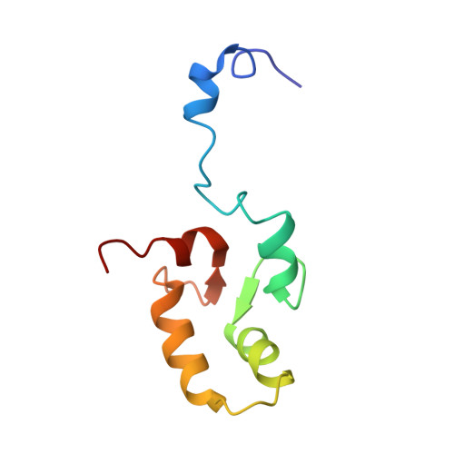
1S6J - PubMed Abstract:
Ca(2+)-dependent protein kinases (CDPKs) are vital Ca(2+)-signaling proteins in plants and protists which have both a kinase domain and a self-contained calcium regulatory calmodulin-like domain (CLD). Despite being very similar to CaM (>40% identity) and sharing the same fold, recent biochemical and structural evidence suggests that the behavior of CLD is distinct from its namesake, calmodulin. In this study, NMR spectroscopy is employed to examine the structure and backbone dynamics of a 168 amino acid Ca(2+)-saturated construct of the CLD (NtH-CLD) in which almost the entire C-terminal domain is exchange broadened and not visible in the NMR spectra. Structural characterization of the N-terminal domain indicates that the first Ca(2+)-binding loop is significantly more open than in a recently reported structure of the CLD complexed with a putative intramolecular binding region (JD) in the CDPK. Backbone dynamics suggest that parts of the third helix exhibit unusually high mobility, and significant exchange, consistent with previous findings that this helix interacts with the C-terminal domain. Dynamics data also show that the "tether" region, consisting of the first 11 amino acids of CLD, is highly mobile and these residues exhibit distinctive beta-type secondary structure, which may help to position the JD and CLD. Finally, the unusual global dynamic behavior of the protein is rationalized on the basis of possible interdomain rearrangements and the highly variable environments of the C- and N-terminal domains.
- Structural Biology Research Group, Department of Biological Sciences, University of Calgary, 2500 University Drive NW, Calgary, Alberta, Canada T2N 1N4.
Organizational Affiliation: