1S60 | pdb_00001s60

Aminoglycoside N-Acetyltransferase AAC(6')-Iy in Complex with CoA and N-terminal His(6)-tag (crystal form 2)


Experimental Data Snapshot

  • Method: X-RAY DIFFRACTION
  • Resolution: 3.00 Å
  • R-Value Free: 
    0.267 (Depositor), 0.263 (DCC) 
  • R-Value Work: 
    0.225 (Depositor), 0.220 (DCC) 
  • R-Value Observed: 
    0.225 (Depositor) 

Starting Model: experimental
View more details

wwPDB Validation 3D Report Full Report

Validation slider image for 1S60

Ligand Structure Quality Assessment 


This is version 1.4 of the entry. See complete history

Literature

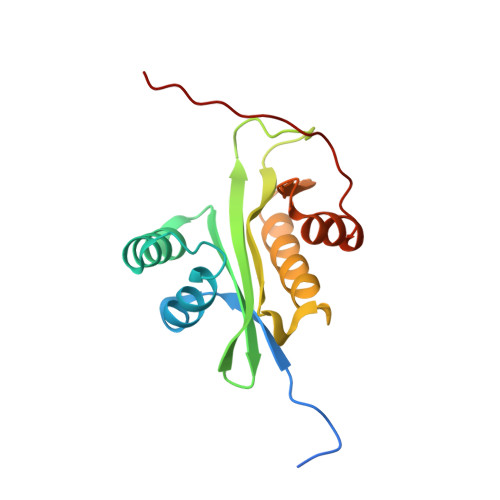
A bacterial acetyltransferase capable of regioselective N-acetylation of antibiotics and histones

Vetting, M.W.Magnet, S.Nieves, E.Roderick, S.L.Blanchard, J.S.

(2004) Chem Biol 11: 565-573

  • DOI: https://doi.org/10.1016/j.chembiol.2004.03.017
  • Primary Citation Related Structures: 
    1S3Z, 1S5K, 1S60

  • PubMed Abstract: 

    The Salmonella enterica chromosomally encoded AAC(6')-Iy has been shown to confer broad aminoglycoside resistance in strains in which the structural gene is expressed. The three-dimensional structures reported place the enzyme in the large Gcn5-related N-acetyltransferase (GNAT) superfamily. The structure of the CoA-ribostamycin ternary complex allows us to propose a chemical mechanism for the reaction, and comparison with the Mycobacterium tuberculosis AAC(2')-CoA-ribostamycin complex allows us to define how regioselectivity of acetylation is achieved. The AAC(6')-Iy dimer is most structurally similar to the Saccharomyces cerevisiae Hpa2-encoded histone acetyltransferase. We demonstrate that AAC(6')-Iy catalyzes both acetyl-CoA-dependent self-alpha-N-acetylation and acetylation of eukaryotic histone proteins and the human histone H3 N-terminal peptide. These structural and catalytic similarities lead us to propose that chromosomally encoded bacterial acetyltransferases, including those functionally identified as aminoglycoside acetyltransferases, are the evolutionary progenitors of the eukaryotic histone acetyltransferases.


  • Organizational Affiliation
    • Department of Biochemistry, Albert Einstein College of Medicine, 1300 Morris Park Avenue, Bronx, NY 10461 USA.

Macromolecule Content 

  • Total Structure Weight: 19.42 kDa 
  • Atom Count: 1,253 
  • Modeled Residue Count: 152 
  • Deposited Residue Count: 165 
  • Unique protein chains: 1

Macromolecules

Find similar proteins by:|  3D Structure
Entity ID: 1
MoleculeChains  Sequence LengthOrganismDetailsImage
aminoglycoside 6'-N-acetyltransferase165Salmonella enterica subsp. enterica serovar EnteritidisMutation(s): 0 
EC: 2.3.1.82
UniProt
Find proteins for Q9R381 (Salmonella enteritidis)
Explore Q9R381 
Go to UniProtKB:  Q9R381
Entity Groups
Sequence Clusters30% Identity50% Identity70% Identity90% Identity95% Identity100% Identity
UniProt GroupQ9R381
Sequence Annotations
Expand
Reference Sequence

Experimental Data & Validation

Experimental Data

  • Method: X-RAY DIFFRACTION
  • Resolution: 3.00 Å
  • R-Value Free:  0.267 (Depositor), 0.263 (DCC) 
  • R-Value Work:  0.225 (Depositor), 0.220 (DCC) 
  • R-Value Observed: 0.225 (Depositor) 
Space Group: P 32 2 1
Unit Cell:
Length ( Å )Angle ( ˚ )
a = 84.6α = 90
b = 84.6β = 90
c = 66.75γ = 120
Software Package:
Software NamePurpose
DENZOdata reduction
SCALEPACKdata scaling
AMoREphasing
CNSrefinement

Structure Validation

View Full Validation Report


Ligand Structure Quality Assessment 


Entry History 

Deposition Data

Revision History  (Full details and data files)

  • Version 1.0: 2004-05-18
    Type: Initial release
  • Version 1.1: 2008-04-29
    Changes: Version format compliance
  • Version 1.2: 2011-07-13
    Changes: Derived calculations, Version format compliance
  • Version 1.3: 2024-02-14
    Changes: Data collection, Database references, Derived calculations
  • Version 1.4: 2024-04-03
    Changes: Refinement description