1S3I | pdb_00001s3i

Crystal structure of the N terminal hydrolase domain of 10-formyltetrahydrofolate dehydrogenase


Experimental Data Snapshot

  • Method: X-RAY DIFFRACTION
  • Resolution: 2.30 Å
  • R-Value Free: 
    0.305 (Depositor), 0.294 (DCC) 
  • R-Value Work: 
    0.242 (Depositor), 0.240 (DCC) 
  • R-Value Observed: 
    0.245 (Depositor) 

wwPDB Validation 3D Report Full Report

Validation slider image for 1S3I

This is version 1.3 of the entry. See complete history

Literature

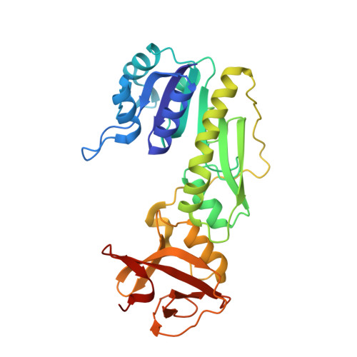
The crystal structure of the hydrolase domain of 10-formyltetrahydrofolate dehydrogenase: mechanism of hydrolysis and its interplay with the dehydrogenase domain.

Chumanevich, A.A.Krupenko, S.A.Davies, C.

(2004) J Biological Chem 279: 14355-14364

  • DOI: https://doi.org/10.1074/jbc.M313934200
  • Primary Citation Related Structures: 
    1S3I

  • PubMed Abstract: 

    10-Formyltetrahydrofolate dehydrogenase (FDH) converts 10-formyltetrahydrofolate, a precursor for nucleotide biosynthesis, to tetrahydrofolate. The protein comprises two functional domains: a hydrolase domain that removes a formyl group from 10-formyltetrahydrofolate and a NADP(+)-dependent dehydrogenase domain that reduces the formyl to carbon dioxide. As a first step toward deciphering the catalytic mechanism of the enzyme, we have determined the crystal structure of the hydrolase domain of FDH from rat, solved to 2.3-A resolution. The structure comprises two domains. As expected, domain 1 shares the same Rossmann fold as the related enzymes, methionyl-tRNA-formyltransferase and glycinamide ribonucleotide formyltransferase, but, unexpectedly, the structural similarity between the amino-terminal domain of 10-formyltetrahydrofolate dehydrogenase and methionyl-tRNA-formyltransferase extends to the C terminus of both proteins. The active site contains a molecule of beta-mercaptoethanol that is positioned between His-106 and Asp-142 and that appears to mimic the formate product. We propose a catalytic mechanism for the hydrolase reaction in which Asp-142 polarizes the catalytic water molecule and His-106 orients the carbonyl group of formyl. The structure also provides clues as to how, in the native enzyme, the hydrolase domain transfers its product to the dehydrogenase domain.


  • Organizational Affiliation
    • Department of Biochemistry and Molecular Biology, Medical University of South Carolina, Charleston, South Carolina 29425, USA.

Macromolecule Content 

  • Total Structure Weight: 34.47 kDa 
  • Atom Count: 2,453 
  • Modeled Residue Count: 307 
  • Deposited Residue Count: 310 
  • Unique protein chains: 1

Macromolecules

Find similar proteins by:|  3D Structure
Entity ID: 1
MoleculeChains  Sequence LengthOrganismDetailsImage
10-formyltetrahydrofolate dehydrogenase310Rattus norvegicusMutation(s): 1 
Gene Names: FTHFD
EC: 1.5.1.6
UniProt
Find proteins for P28037 (Rattus norvegicus)
Explore P28037 
Go to UniProtKB:  P28037
Entity Groups
Sequence Clusters30% Identity50% Identity70% Identity90% Identity95% Identity100% Identity
UniProt GroupP28037
Sequence Annotations
Expand
Reference Sequence

Experimental Data & Validation

Experimental Data

  • Method: X-RAY DIFFRACTION
  • Resolution: 2.30 Å
  • R-Value Free:  0.305 (Depositor), 0.294 (DCC) 
  • R-Value Work:  0.242 (Depositor), 0.240 (DCC) 
  • R-Value Observed: 0.245 (Depositor) 
Space Group: P 21 21 2
Unit Cell:
Length ( Å )Angle ( ˚ )
a = 100α = 90
b = 64.63β = 90
c = 64.59γ = 90
Software Package:
Software NamePurpose
REFMACrefinement
CrystalCleardata reduction
d*TREKdata scaling
PHASESphasing

Structure Validation

View Full Validation Report


Entry History 

Deposition Data

Revision History  (Full details and data files)

  • Version 1.0: 2004-01-27
    Type: Initial release
  • Version 1.1: 2008-04-29
    Changes: Version format compliance
  • Version 1.2: 2011-07-13
    Changes: Version format compliance
  • Version 1.3: 2021-10-27
    Changes: Database references, Derived calculations