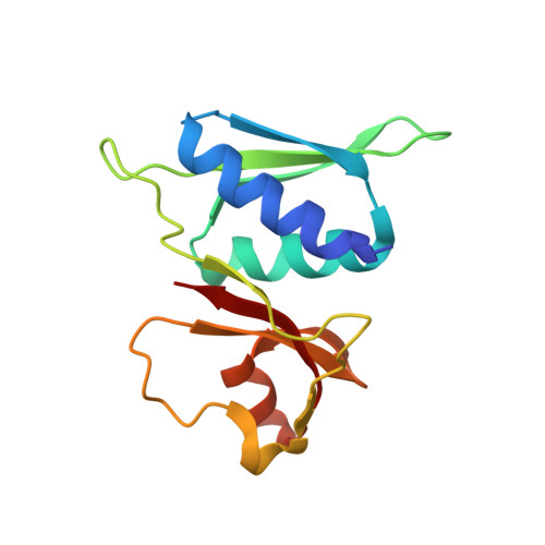
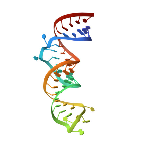
1S03

The Structure of a Ribosomal Protein S8/spc Operon mRNA Complex


Experimental Data Snapshot

  • Method: X-RAY DIFFRACTION
  • Resolution: 2.70 Å
  • R-Value Free: 0.273 
  • R-Value Work: 0.237 
  • R-Value Observed: 0.237 

Starting Model: experimental
View more details

wwPDB Validation   3D Report Full Report


This is version 1.3 of the entry. See complete history


Literature

The structure of a ribosomal protein S8/spc operon mRNA complex.

Merianos, H.J.Wang, J.Moore, P.B.

(2004) RNA 10: 954-964

  • DOI: https://doi.org/10.1261/rna.7030704
  • Primary Citation of Related Structures:  
    1S03

  • PubMed Abstract: 

    In bacteria, translation of all the ribosomal protein cistrons in the spc operon mRNA is repressed by the binding of the product of one of them, S8, to an internal sequence at the 5' end of the L5 cistron. The way in which the first two genes of the spc operon are regulated, retroregulation, is mechanistically distinct from translational repression by S8 of the genes from L5 onward. A 2.8 A resolution crystal structure has been obtained of Escherichia coli S8 bound to this site. Despite sequence differences, the structure of this complex is almost identical to that of the S8/helix 21 complex seen in the small ribosomal subunit, consistent with the hypothesis that autogenous regulation of ribosomal protein synthesis results from conformational similarities between mRNAs and rRNAs. S8 binding must repress the translation of its own mRNA by inhibiting the formation of a ribosomal initiation complex at the start of the L5 cistron.


  • Organizational Affiliation

    Department of Chemistry, Yale University, New Haven, Connecticut 06520, USA.


Macromolecules

Find similar proteins by:  (by identity cutoff)  |  3D Structure
Entity ID: 2
MoleculeChains Sequence LengthOrganismDetailsImage
30S ribosomal protein S8C [auth H],
D [auth G]
129Escherichia coliMutation(s): 0 
UniProt
Find proteins for P0A7W7 (Escherichia coli (strain K12))
Explore P0A7W7 
Go to UniProtKB:  P0A7W7
Entity Groups  
Sequence Clusters30% Identity50% Identity70% Identity90% Identity95% Identity100% Identity
UniProt GroupP0A7W7
Sequence Annotations
Expand
  • Reference Sequence
Find similar nucleic acids by:  (by identity cutoff)  |  3D Structure
Entity ID: 1
MoleculeChains LengthOrganismImage
47-MER
A, B
47N/A
Sequence Annotations
Expand
  • Reference Sequence
Small Molecules
Ligands 1 Unique
IDChains Name / Formula / InChI Key2D Diagram3D Interactions
ZN
Query on ZN

Download Ideal Coordinates CCD File 
E [auth A]
F [auth A]
G [auth A]
H [auth A]
I [auth A]
E [auth A],
F [auth A],
G [auth A],
H [auth A],
I [auth A],
J [auth A],
K [auth A],
L [auth A],
M [auth B],
N [auth B],
O [auth B],
P [auth B],
Q [auth B],
R [auth B],
S [auth H],
T [auth G],
U [auth G]
ZINC ION
Zn
PTFCDOFLOPIGGS-UHFFFAOYSA-N
Experimental Data & Validation

Experimental Data

  • Method: X-RAY DIFFRACTION
  • Resolution: 2.70 Å
  • R-Value Free: 0.273 
  • R-Value Work: 0.237 
  • R-Value Observed: 0.237 
  • Space Group: P 32 2 1
Unit Cell:
Length ( Å )Angle ( ˚ )
a = 61.22α = 90
b = 61.22β = 90
c = 332.52γ = 120
Software Package:
Software NamePurpose
REFMACrefinement
HKL-2000data reduction
SCALEPACKdata scaling
AMoREphasing

Structure Validation

View Full Validation Report


Entry History 

Deposition Data

Revision History  (Full details and data files)

  • Version 1.0: 2004-05-25
    Type: Initial release
  • Version 1.1: 2008-04-29
    Changes: Version format compliance
  • Version 1.2: 2011-07-13
    Changes: Advisory, Version format compliance
  • Version 1.3: 2023-08-23
    Changes: Data collection, Database references, Derived calculations, Refinement description