DNA A-tract bending in three dimensions: Solving the dA4T4 vs. dT4A4 conundrum.
Stefl, R., Wu, H., Ravindranathan, S., Sklenar, V., Feigon, J.(2004) Proc Natl Acad Sci U S A 101: 1177-1182
- PubMed: 14739342 Search on PubMedSearch on PubMed Central
- DOI: https://doi.org/10.1073/pnas.0308143100
- Primary Citation Related Structures:
1RVH, 1RVI - PubMed Abstract:
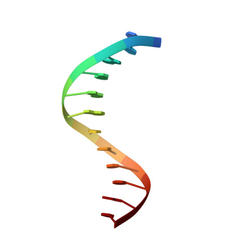
DNA A-tracts have been defined as four or more consecutive A.T base pairs without a TpA step. When inserted in phase with the DNA helical repeat, bending is manifested macroscopically as anomalous migration on polyacrylamide gels, first observed >20 years ago. An unsolved conundrum is why DNA containing in-phase A-tract repeats of A(4)T(4) are bent, whereas T(4)A(4) is straight. We have determined the solution structures of the DNA duplexes formed by d(GCAAAATTTTGC) [A4T4] and d(CGTTTTAAAACG) [T4A4] with NH(4)(+) counterions by using NMR spectroscopy, including refinement with residual dipolar couplings. Analysis of the structures shows that the ApT step has a large negative roll, resulting in a local bend toward the minor groove, whereas the TpA step has a positive roll and locally bends toward the major groove. For A4T4, this bend is nearly in phase with bends at the two A-tract junctions, resulting in an overall bend toward the minor groove of the A-tract, whereas for T4A4, the bends oppose each other, resulting in a relatively straight helix. NMR-based structural modeling of d(CAAAATTTTG)(15) and d(GTTTTAAAAC)(15) reveals that the former forms a left-handed superhelix with a diameter of approximately 110 A and pitch of 80 A, similar to DNA in the nucleosome, whereas the latter has a gentle writhe with a pitch of >250 A and diameter of approximately 50 A. Results of gel electrophoretic mobility studies are consistent with the higher-order structure of the DNA and furthermore depend on the nature of the monovalent cation present in the running buffer.
- National Center for Biomolecular Research, NMR Laboratory, Faculty of Science, Masaryk University, Kotlárská 2, CZ-611 37 Brno, Czech Republic.
Organizational Affiliation: