1RPT | pdb_00001rpt

CRYSTAL STRUCTURES OF RAT ACID PHOSPHATASE COMPLEXED WITH THE TRANSITIONS STATE ANALOGS VANADATE AND MOLYBDATE: IMPLICATIONS FOR THE REACTION MECHANISM


Experimental Data Snapshot

  • Method: X-RAY DIFFRACTION
  • Resolution: 3.00 Å
  • R-Value Work: 
    0.215 (Depositor) 
  • R-Value Observed: 
    0.215 (Depositor) 

wwPDB Validation 3D Report Full Report

Validation slider image for 1RPT

This is version 2.1 of the entry. See complete history

Literature

Crystal structures of rat acid phosphatase complexed with the transition-state analogs vanadate and molybdate. Implications for the reaction mechanism.

Lindqvist, Y.Schneider, G.Vihko, P.

(1994) Eur J Biochem 221: 139-142

  • DOI: https://doi.org/10.1111/j.1432-1033.1994.tb18722.x
  • Primary Citation Related Structures: 
    1RPT

  • PubMed Abstract: 

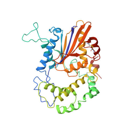
    The three-dimensional structures of complexes of recombinant rat prostatic acid phosphatase with the transition-state analogs vanadate and molybdate were determined to 0.3-nm resolution using protein crystallographic methods. The overall structure of the enzyme remains unchanged upon binding of the metal oxyanions; only local conformational differences in the positions of some side chains at the active site were found. The metal oxyanions bind in an identical fashion at the active site with trigonal bipyramidal coordination geometry. The metal ion is within coordination distance of the His12 side chain which is located at one of the axial positions. The three equatorial oxygen atoms interact with the conserved residues Arg11, Arg15, Arg79 and His257. Within hydrogen-bonding distance of the axial oxygen atom is the side chain of the conserved residue Asp258. The implications of these results for the catalytic mechanism of acid phosphatase are discussed.


  • Organizational Affiliation
    • Department of Molecular Biology, Swedish University of Agricultural Sciences, Uppsala.

Macromolecule Content 

  • Total Structure Weight: 40.61 kDa 
  • Atom Count: 2,852 
  • Modeled Residue Count: 342 
  • Deposited Residue Count: 342 
  • Unique protein chains: 1

Macromolecules

Find similar proteins by:|  3D Structure
Entity ID: 1
MoleculeChains  Sequence LengthOrganismDetailsImage
PROSTATIC ACID PHOSPHATASE342Rattus norvegicusMutation(s): 0 
EC: 3.1.3.2 (PDB Primary Data), 3.1.3.5 (UniProt), 3.1.3.48 (UniProt)
UniProt
Find proteins for P20646 (Rattus norvegicus)
Explore P20646 
Go to UniProtKB:  P20646
Entity Groups
Sequence Clusters30% Identity50% Identity70% Identity90% Identity95% Identity100% Identity
UniProt GroupP20646
Glycosylation
Glycosylation Sites: 2Go to GlyGen: P20646-1
Sequence Annotations
Expand
Reference Sequence

Oligosaccharides

Help  
Entity ID: 2
MoleculeChains Length2D Diagram GlycosylationD Interactions
beta-D-mannopyranose-(1-4)-2-acetamido-2-deoxy-beta-D-glucopyranose-(1-4)-2-acetamido-2-deoxy-beta-D-glucopyranose
B
3N-Glycosylation
Glycosylation Resources
GlyTouCan: G15407YE
GlyCosmos: G15407YE
GlyGen: G15407YE

Small Molecules

Ligands 2 Unique
IDChains Name / Formula / InChI Key2D Diagram3D Interactions
NAG

Query on NAG


Download:Ideal Coordinates CCD File
C [auth A]2-acetamido-2-deoxy-beta-D-glucopyranose
C8 H15 N O6
OVRNDRQMDRJTHS-FMDGEEDCSA-N
VO4

Query on VO4


Download:Ideal Coordinates CCD File
D [auth A]VANADATE ION
O4 V
LSGOVYNHVSXFFJ-UHFFFAOYSA-N

Experimental Data & Validation

Experimental Data

  • Method: X-RAY DIFFRACTION
  • Resolution: 3.00 Å
  • R-Value Work:  0.215 (Depositor) 
  • R-Value Observed: 0.215 (Depositor) 
Space Group: P 32 2 1
Unit Cell:
Length ( Å )Angle ( ˚ )
a = 89.4α = 90
b = 89.4β = 90
c = 152γ = 120
Software Package:
Software NamePurpose
X-PLORmodel building
X-PLORrefinement
X-PLORphasing

Structure Validation

View Full Validation Report


Entry History 

Deposition Data

Revision History  (Full details and data files)

  • Version 1.0: 1994-05-31
    Type: Initial release
  • Version 1.1: 2008-03-24
    Changes: Version format compliance
  • Version 1.2: 2011-07-13
    Changes: Non-polymer description, Version format compliance
  • Version 2.0: 2020-07-29
    Type: Remediation
    Reason: Carbohydrate remediation
    Changes: Atomic model, Data collection, Database references, Derived calculations, Other, Structure summary
  • Version 2.1: 2024-10-16
    Changes: Data collection, Database references, Structure summary