1RKI | pdb_00001rki

Structure of pag5_736 from P. aerophilum with three disulphide bonds


Experimental Data Snapshot

  • Method: X-RAY DIFFRACTION
  • Resolution: 1.60 Å
  • R-Value Free: 
    0.235 (Depositor) 
  • R-Value Work: 
    0.184 (Depositor) 
  • R-Value Observed: 
    0.187 (Depositor) 

wwPDB Validation 3D Report Full Report

Validation slider image for 1RKI

This is version 1.3 of the entry. See complete history

Literature

The Genomics of Disulfide Bonding and Protein Stabilization in Thermophiles.

Beeby, M.Ryttersgaard, C.Boutz, D.R.Perry, L.J.Yeates, T.O.

(2005) PLoS Biol 3: e309-e309

  • DOI: https://doi.org/10.1371/journal.pbio.0030309
  • Primary Citation Related Structures: 
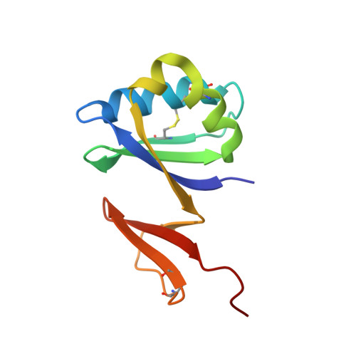
    1RKI

  • PubMed Abstract: 

    Thermophilic organisms flourish in varied high-temperature environmental niches that are deadly to other organisms. Recently, genomic evidence has implicated a critical role for disulfide bonds in the structural stabilization of intracellular proteins from certain of these organisms, contrary to the conventional view that structural disulfide bonds are exclusively extracellular. Here both computational and structural data are presented to explore the occurrence of disulfide bonds as a protein-stabilization method across many thermophilic prokaryotes. Based on computational studies, disulfide-bond richness is found to be widespread, with thermophiles containing the highest levels. Interestingly, only a distinct subset of thermophiles exhibit this property. A computational search for proteins matching this target phylogenetic profile singles out a specific protein, known as protein disulfide oxidoreductase, as a potential key player in thermophilic intracellular disulfide-bond formation. Finally, biochemical support in the form of a new crystal structure of a thermophilic protein with three disulfide bonds is presented together with a survey of known structures from the literature. Together, the results provide insight into biochemical specialization and the diversity of methods employed by organisms to stabilize their proteins in exotic environments. The findings also motivate continued efforts to sequence genomes from divergent organisms.


  • Organizational Affiliation
    • UCLA-DOE Institute for Genomics and Proteomics, University of California, Los Angeles, California, USA.

Macromolecule Content 

  • Total Structure Weight: 24.56 kDa 
  • Atom Count: 1,759 
  • Modeled Residue Count: 198 
  • Deposited Residue Count: 204 
  • Unique protein chains: 1

Macromolecules

Find similar proteins by:|  3D Structure
Entity ID: 1
MoleculeChains  Sequence LengthOrganismDetailsImage
hypothetical protein
A, B
102Pyrobaculum aerophilumMutation(s): 0 
UniProt
Find proteins for Q8ZYK2 (Pyrobaculum aerophilum (strain ATCC 51768 / DSM 7523 / JCM 9630 / CIP 104966 / NBRC 100827 / IM2))
Explore Q8ZYK2 
Go to UniProtKB:  Q8ZYK2
Entity Groups
Sequence Clusters30% Identity50% Identity70% Identity90% Identity95% Identity100% Identity
UniProt GroupQ8ZYK2
Sequence Annotations
Expand
Reference Sequence

Small Molecules

Experimental Data & Validation

Experimental Data

  • Method: X-RAY DIFFRACTION
  • Resolution: 1.60 Å
  • R-Value Free:  0.235 (Depositor) 
  • R-Value Work:  0.184 (Depositor) 
  • R-Value Observed: 0.187 (Depositor) 
Space Group: C 1 2 1
Unit Cell:
Length ( Å )Angle ( ˚ )
a = 105.483α = 90
b = 43.717β = 113.24
c = 52.717γ = 90
Software Package:
Software NamePurpose
REFMACrefinement
DENZOdata reduction
SCALEPACKdata scaling
SHELXDphasing
MLPHAREphasing
DMphasing
CNSrefinement

Structure Validation

View Full Validation Report


Entry History 

Deposition Data

Revision History  (Full details and data files)

  • Version 1.0: 2005-01-18
    Type: Initial release
  • Version 1.1: 2008-04-29
    Changes: Version format compliance
  • Version 1.2: 2011-07-13
    Changes: Advisory, Refinement description, Version format compliance
  • Version 1.3: 2024-10-30
    Changes: Data collection, Database references, Derived calculations, Structure summary