1RIN

X-RAY CRYSTAL STRUCTURE OF A PEA LECTIN-TRIMANNOSIDE COMPLEX AT 2.6 ANGSTROMS RESOLUTION


Experimental Data Snapshot

  • Method: X-RAY DIFFRACTION
  • Resolution: 2.60 Å
  • R-Value Work: 0.183 
  • R-Value Observed: 0.183 

wwPDB Validation   3D Report Full Report


This is version 1.4 of the entry. See complete history


Literature

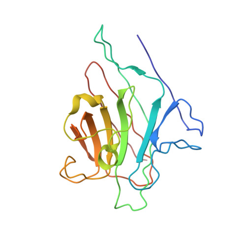
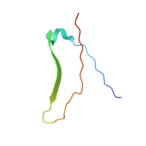
X-ray crystal structure of a pea lectin-trimannoside complex at 2.6 A resolution.

Rini, J.M.Hardman, K.D.Einspahr, H.Suddath, F.L.Carver, J.P.

(1993) J Biol Chem 268: 10126-10132

  • DOI: https://doi.org/10.2210/pdb1rin/pdb
  • Primary Citation of Related Structures:  
    1RIN

  • PubMed Abstract: 

    The x-ray crystal structure of pea lectin, in complex with a methyl glycoside of the N-linked-type oligosaccharide trimannosyl core, methyl 3,6-di-O-(alpha-D-mannopyranosyl)-alpha-D-mannopyranoside, has been solved by molecular replacement and refined at 2.6-A resolution. The R factor is 0.183 for all data in the 8.0 to 2.6 A resolution range with an average atomic temperature factor of 26.1 A2. Strong electron density for a single mannose residue is found in the monosaccharide-binding site suggesting that the trisaccharide binds primarily through one of the terminal alpha-linked mannose residues. The complex is stabilized by hydrogen bonds involving the protein residues Asp-81, Gly-99, Asn-125, Ala-217, and Glu-218, and the carbohydrate oxygen atoms O3, O4, O5, and O6. In addition, the carbohydrate makes van der Waals contacts with the protein, involving Phe-123 in particular. These interactions are very similar to those found in the monosaccharide complexes with concanavalin A and isolectin 1 of Lathyrus ochrus, confirming the structural relatedness of this family of proteins. Comparison of the pea lectin complex with the unliganded pea lectin and concanavalin A structures indicates differences in the conformation and water structure of the unliganded binding sites of these two proteins. Furthermore, a correlation between the position of the carbohydrate oxygen atoms in the complex and the bound water molecules in the unliganded binding sites is found. Binding of the trimannose core through a single terminal monosaccharide residue strongly argues that an additional fucose-binding site is responsible for the high affinity pea lectin-oligosaccharide interactions.


  • Organizational Affiliation

    Department of Molecular and Medical Genetics, University of Toronto, Ontario, Canada.


Macromolecules
Find similar proteins by:  (by identity cutoff)  |  3D Structure
Entity ID: 1
MoleculeChains Sequence LengthOrganismDetailsImage
PEA LECTIN
A, C
180Pisum sativumMutation(s): 0 
UniProt
Find proteins for P02867 (Pisum sativum)
Explore P02867 
Go to UniProtKB:  P02867
Entity Groups  
Sequence Clusters30% Identity50% Identity70% Identity90% Identity95% Identity100% Identity
UniProt GroupP02867
Sequence Annotations
Expand
  • Reference Sequence
Find similar proteins by:  (by identity cutoff)  |  3D Structure
Entity ID: 2
MoleculeChains Sequence LengthOrganismDetailsImage
PEA LECTIN
B, D
49Pisum sativumMutation(s): 0 
UniProt
Find proteins for P02867 (Pisum sativum)
Explore P02867 
Go to UniProtKB:  P02867
Entity Groups  
Sequence Clusters30% Identity50% Identity70% Identity90% Identity95% Identity100% Identity
UniProt GroupP02867
Sequence Annotations
Expand
  • Reference Sequence
Small Molecules
Binding Affinity Annotations 
IDSourceBinding Affinity
MAN PDBBind:  1RIN Kd: 7.00e+5 (nM) from 1 assay(s)
Experimental Data & Validation

Experimental Data

  • Method: X-RAY DIFFRACTION
  • Resolution: 2.60 Å
  • R-Value Work: 0.183 
  • R-Value Observed: 0.183 
  • Space Group: P 21 21 21
Unit Cell:
Length ( Å )Angle ( ˚ )
a = 64.3α = 90
b = 73.4β = 90
c = 108.5γ = 90
Software Package:
Software NamePurpose
X-PLORmodel building
X-PLORrefinement
X-PLORphasing

Structure Validation

View Full Validation Report


Entry History 

Deposition Data

Revision History  (Full details and data files)

  • Version 1.0: 1993-10-31
    Type: Initial release
  • Version 1.1: 2008-03-24
    Changes: Version format compliance
  • Version 1.2: 2011-07-13
    Changes: Derived calculations, Version format compliance
  • Version 1.3: 2020-07-29
    Type: Remediation
    Reason: Carbohydrate remediation
    Changes: Data collection, Database references, Derived calculations, Other, Structure summary
  • Version 1.4: 2024-02-14
    Changes: Data collection, Database references, Structure summary