1RIF | pdb_00001rif

Crystal structure of the UvsW helicase from Bacteriophage T4


Experimental Data Snapshot

  • Method: X-RAY DIFFRACTION
  • Resolution: 2.00 Å
  • R-Value Free: 
    0.246 (Depositor) 
  • R-Value Work: 
    0.218 (Depositor) 
  • R-Value Observed: 
    0.221 (Depositor) 

wwPDB Validation 3D Report Full Report

Validation slider image for 1RIF

This is version 1.4 of the entry. See complete history

Literature

The Crystal Structure of the UvsW Helicase from Bacteriophage T4.

Sickmier, E.A.Kreuzer, K.N.White, S.W.

(2004) Structure 12: 583-592

  • DOI: https://doi.org/10.1016/j.str.2004.02.016
  • Primary Citation Related Structures: 
    1RIF

  • PubMed Abstract: 

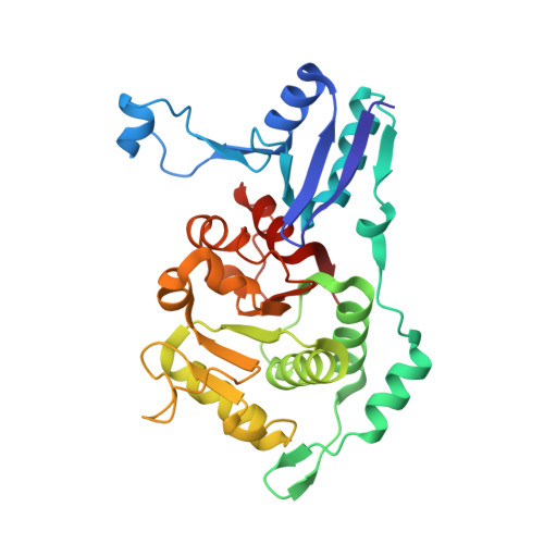
    In bacteriophage T4, the WXY system repairs DNA damage by a process that involves homologous recombination. This system comprises three proteins, the RecA-like recombination protein UvsX, a recombination mediator protein UvsY, and a helicase UvsW. Here we report the 2.0 A resolution crystal structure of the N-terminal two domains of the UvsW helicase (UvsWNF; residues 1-282). The structure reveals a typical helicase RecA-like domain linked to a small N-terminal alpha/beta domain that likely binds the nucleic acid substrate. The missing C-terminal portion of UvsW almost certainly corresponds to the second RecA-like domain typically found in monomeric helicases. The putative substrate binding domain is unique within the known helicase structures, and it resembles the novel "double-wing" DNA binding domain from the phage T4 MotA transcription factor that mediates the expression of T4 middle genes. The functional implications of this homology for the role of UvsW in T4 DNA metabolism are discussed.


  • Organizational Affiliation
    • Department of Biochemistry, Duke University Medical Center, Durham, NC 27710, USA.

Macromolecule Content 

  • Total Structure Weight: 66.55 kDa 
  • Atom Count: 4,891 
  • Modeled Residue Count: 564 
  • Deposited Residue Count: 564 
  • Unique protein chains: 1

Macromolecules

Find similar proteins by:|  3D Structure
Entity ID: 1
MoleculeChains  Sequence LengthOrganismDetailsImage
DNA helicase uvsW
A, B
282Tequatrovirus T4Mutation(s): 1 
Gene Names: UVSWDAR
EC: 5.6.2.4
UniProt
Find proteins for P0DXF1 (Enterobacteria phage T4)
Explore P0DXF1 
Go to UniProtKB:  P0DXF1
Entity Groups
Sequence Clusters30% Identity50% Identity70% Identity90% Identity95% Identity100% Identity
UniProt GroupP0DXF1
Sequence Annotations
Expand
Reference Sequence

Experimental Data & Validation

Experimental Data

  • Method: X-RAY DIFFRACTION
  • Resolution: 2.00 Å
  • R-Value Free:  0.246 (Depositor) 
  • R-Value Work:  0.218 (Depositor) 
  • R-Value Observed: 0.221 (Depositor) 
Space Group: P 1 21 1
Unit Cell:
Length ( Å )Angle ( ˚ )
a = 42.739α = 90
b = 95.181β = 95.4
c = 86.278γ = 90
Software Package:
Software NamePurpose
HKL-2000data collection
SCALEPACKdata scaling
CNSrefinement
HKL-2000data reduction
CNSphasing

Structure Validation

View Full Validation Report


Entry History 

Deposition Data

Revision History  (Full details and data files)

  • Version 1.0: 2003-11-25
    Type: Initial release
  • Version 1.1: 2008-04-29
    Changes: Version format compliance
  • Version 1.2: 2011-07-13
    Changes: Version format compliance
  • Version 1.3: 2021-10-27
    Changes: Database references, Derived calculations
  • Version 1.4: 2024-02-14
    Changes: Data collection