1RI9 | pdb_00001ri9

Structure of a helically extended SH3 domain of the T cell adapter protein ADAP


Experimental Data Snapshot

  • Method: SOLUTION NMR
  • Conformers Calculated: 51 
  • Conformers Submitted: 20 
  • Selection Criteria: The submitted conformer models are the 20 structures with the lowest energy. 

wwPDB Validation 3D Report Full Report

Validation slider image for 1RI9

This is version 1.5 of the entry. See complete history

Literature

Structure of a Helically Extended SH3 Domain of the T Cell Adapter Protein ADAP.

Heuer, K.Kofler, M.Langdon, G.Thiemke, K.Freund, C.

(2004) Structure 12: 603-610

  • DOI: https://doi.org/10.1016/j.str.2004.02.021
  • Primary Citation Related Structures: 
    1RI9

  • PubMed Abstract: 

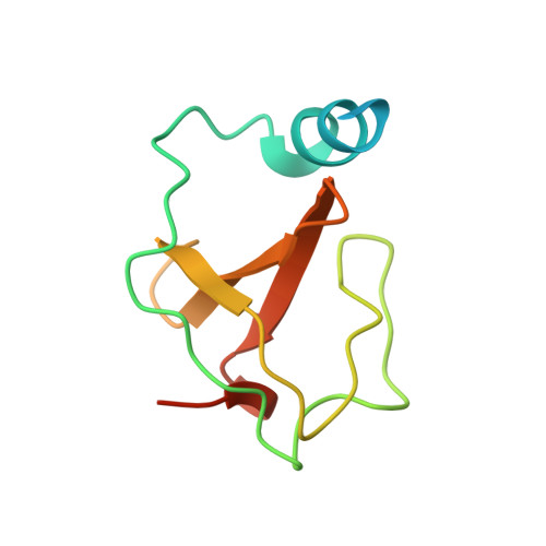
    The adapter protein ADAP (FYB/SLAP-130) provides a critical link between T cell receptor (TCR) signaling and cell adhesion via the activation of integrins. The C-terminal 70 residues of ADAP show homology to SH3 domains; however, conserved residues of the fold are absent. An alignment and annotation of this domain has therefore been elusive. We have solved the three-dimensional structure of the ADAP C-terminal domain by NMR spectroscopy and show that it represents an altered SH3 domain fold. An N-terminal, amphipathic helix makes extensive contacts to residues of the regular SH3 domain fold, and thereby a composite surface with unusual surface properties is created. We propose this SH3 domain variant to be classified as a helically extended SH3 domain (hSH3 domain) and show that the ADAP-hSH3 domain can no longer bind conventional proline-rich peptides.


  • Organizational Affiliation
    • Protein Engineering Group, Forschungsinstitut für Molekulare Pharmakologie and Freie Universität Berlin, Robert-Rössle-Strasse 10, 13125 Berlin, Germany.

Macromolecule Content 

  • Total Structure Weight: 11.9 kDa 
  • Atom Count: 634 
  • Modeled Residue Count: 77 
  • Deposited Residue Count: 102 
  • Unique protein chains: 1

Macromolecules

Find similar proteins by:|  3D Structure
Entity ID: 1
MoleculeChains  Sequence LengthOrganismDetailsImage
FYN-binding protein102Homo sapiensMutation(s): 0 
Gene Names: FYBSLAP130
UniProt & NIH Common Fund Data Resources
Find proteins for O15117 (Homo sapiens)
Explore O15117 
Go to UniProtKB:  O15117
PHAROS:  O15117
GTEx:  ENSG00000082074 
Entity Groups
Sequence Clusters30% Identity50% Identity70% Identity90% Identity95% Identity100% Identity
UniProt GroupO15117
Sequence Annotations
Expand
Reference Sequence

Experimental Data & Validation

Experimental Data

  • Method: SOLUTION NMR
  • Conformers Calculated: 51 
  • Conformers Submitted: 20 
  • Selection Criteria: The submitted conformer models are the 20 structures with the lowest energy. 

Structure Validation

View Full Validation Report


Entry History 

Deposition Data

Revision History  (Full details and data files)

  • Version 1.0: 2004-04-20
    Type: Initial release
  • Version 1.1: 2008-04-29
    Changes: Version format compliance
  • Version 1.2: 2011-07-13
    Changes: Version format compliance
  • Version 1.3: 2020-02-05
    Changes: Data collection, Database references, Derived calculations, Other
  • Version 1.4: 2023-06-14
    Changes: Database references, Other
  • Version 1.5: 2024-05-08
    Changes: Data collection, Database references