1RFY | pdb_00001rfy

Crystal Structure of Quorum-Sensing Antiactivator TraM


Experimental Data Snapshot

  • Method: X-RAY DIFFRACTION
  • Resolution: 1.60 Å
  • R-Value Free: 
    0.222 (Depositor), 0.222 (DCC) 
  • R-Value Work: 
    0.202 (Depositor), 0.202 (DCC) 
  • R-Value Observed: 
    0.202 (Depositor) 

wwPDB Validation 3D Report Full Report

Validation slider image for 1RFY

This is version 1.4 of the entry. See complete history

Literature

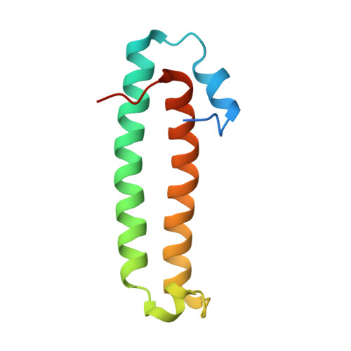
Quorum-sensing antiactivator TraM forms a dimer that dissociates to inhibit TraR

Chen, G.Malenkos, J.W.Cha, M.R.Fuqua, C.Chen, L.

(2004) Mol Microbiol 52: 1641-1651

  • DOI: https://doi.org/10.1111/j.1365-2958.2004.04110.x
  • Primary Citation Related Structures: 
    1RFY

  • PubMed Abstract: 

    The quorum-sensing transcriptional activator TraR of Agrobacterium tumefaciens, which controls the replication and conjugal transfer of the tumour-inducing (Ti) virulence plasmid, is inhibited by the TraM antiactivator. The crystal structure of TraM reveals this protein to form a homodimer in which the monomer primarily consists of two long coiled alpha-helices, and one of the helices from each monomer also bundles to form the dimeric interface. The importance of dimerization is addressed by mutational studies in which disruption of the hydrophobic dimer interface leads to aggregation of TraM. Biochemical studies confirm that TraM exists as a homodimer in solution in equilibrium with the monomeric form, and also establish that the TraM-TraR complex is a heterodimer. Thus, the TraM homodimer undergoes dissociation in forming the antiactivation complex. Combined with the structure of TraR (Zhang et al., 2002, Nature 417: 971-974; Vannini et al., 2002, EMBO J 21: 4393-4401), our structural analysis suggests overlapping interactive surfaces in homodimeric TraM with those in the TraM-TraR complex and a mechanism for TraM inhibition on TraR.


  • Organizational Affiliation
    • Department of Biology, Indiana University, 915 E. 3rd St., Bloomington, IN 47405, USA.

Macromolecule Content 

  • Total Structure Weight: 22.73 kDa 
  • Atom Count: 1,579 
  • Modeled Residue Count: 177 
  • Deposited Residue Count: 204 
  • Unique protein chains: 1

Macromolecules

Find similar proteins by:|  3D Structure
Entity ID: 1
MoleculeChains  Sequence LengthOrganismDetailsImage
Transcriptional repressor traM
A, B
102Agrobacterium tumefaciensMutation(s): 0 
Gene Names: traM
UniProt
Find proteins for Q57471 (Agrobacterium tumefaciens)
Explore Q57471 
Go to UniProtKB:  Q57471
Entity Groups
Sequence Clusters30% Identity50% Identity70% Identity90% Identity95% Identity100% Identity
UniProt GroupQ57471
Sequence Annotations
Expand
Reference Sequence

Experimental Data & Validation

Experimental Data

  • Method: X-RAY DIFFRACTION
  • Resolution: 1.60 Å
  • R-Value Free:  0.222 (Depositor), 0.222 (DCC) 
  • R-Value Work:  0.202 (Depositor), 0.202 (DCC) 
  • R-Value Observed: 0.202 (Depositor) 
Space Group: C 2 2 2
Unit Cell:
Length ( Å )Angle ( ˚ )
a = 78.23α = 90
b = 85.75β = 90
c = 67.02γ = 90
Software Package:
Software NamePurpose
DENZOdata reduction
SCALEPACKdata scaling
SnBphasing
SOLVEphasing
CNSrefinement

Structure Validation

View Full Validation Report


Entry History 

Deposition Data

Revision History  (Full details and data files)

  • Version 1.0: 2004-11-23
    Type: Initial release
  • Version 1.1: 2008-04-29
    Changes: Version format compliance
  • Version 1.2: 2011-07-13
    Changes: Derived calculations, Version format compliance
  • Version 1.3: 2017-10-11
    Changes: Refinement description
  • Version 1.4: 2024-02-14
    Changes: Data collection, Database references