1RFX | pdb_00001rfx

Crystal Structure of resisitin


Experimental Data Snapshot

  • Method: X-RAY DIFFRACTION
  • Resolution: 2.00 Å
  • R-Value Free: 
    0.216 (Depositor), 0.222 (DCC) 
  • R-Value Work: 
    0.177 (Depositor), 0.185 (DCC) 
  • R-Value Observed: 
    0.179 (Depositor) 

wwPDB Validation 3D Report Full Report

Validation slider image for 1RFX

This is version 1.4 of the entry. See complete history

Literature

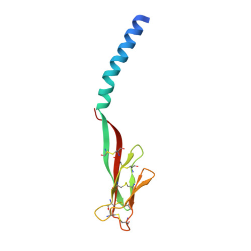
Disulfide-dependent multimeric assembly of resistin family hormones

Patel, S.D.Rajala, M.W.Rossetti, L.Scherer, P.E.Shapiro, L.

(2004) Science 304: 1154-1158

  • DOI: https://doi.org/10.1126/science.1093466
  • Primary Citation Related Structures: 
    1RFX, 1RGX, 1RH7

  • PubMed Abstract: 

    Resistin, founding member of the resistin-like molecule (RELM) hormone family, is secreted selectively from adipocytes and induces liver-specific antagonism of insulin action, thus providing a potential molecular link between obesity and diabetes. Crystal structures of resistin and RELMbeta reveal an unusual multimeric structure. Each protomer comprises a carboxy-terminal disulfide-rich beta-sandwich "head" domain and an amino-terminal alpha-helical "tail" segment. The alpha-helical segments associate to form three-stranded coiled coils, and surface-exposed interchain disulfide linkages mediate the formation of tail-to-tail hexamers. Analysis of serum samples shows that resistin circulates in two distinct assembly states, likely corresponding to hexamers and trimers. Infusion of a resistin mutant, lacking the intertrimer disulfide bonds, in pancreatic-insulin clamp studies reveals substantially more potent effects on hepatic insulin sensitivity than those observed with wild-type resistin. This result suggests that processing of the intertrimer disulfide bonds may reflect an obligatory step toward activation.


  • Organizational Affiliation
    • Department of Biochemistry and Molecular Biophysics, Columbia University, New York, NY 10032, USA.

Macromolecule Content 

  • Total Structure Weight: 31.61 kDa 
  • Atom Count: 2,279 
  • Modeled Residue Count: 267 
  • Deposited Residue Count: 282 
  • Unique protein chains: 1

Macromolecules

Find similar proteins by:|  3D Structure
Entity ID: 1
MoleculeChains  Sequence LengthOrganismDetailsImage
Resistin
A, B, C
94Mus musculusMutation(s): 0 
Gene Names: RETN
Membrane Entity: Yes 
UniProt
Find proteins for Q99P87 (Mus musculus)
Explore Q99P87 
Go to UniProtKB:  Q99P87
Entity Groups
Sequence Clusters30% Identity50% Identity70% Identity90% Identity95% Identity100% Identity
UniProt GroupQ99P87
Sequence Annotations
Expand
Reference Sequence

Small Molecules

Ligands 4 Unique
IDChains Name / Formula / InChI Key2D Diagram3D Interactions
PGE

Query on PGE


Download:Ideal Coordinates CCD File
R [auth B]TRIETHYLENE GLYCOL
C6 H14 O4
ZIBGPFATKBEMQZ-UHFFFAOYSA-N
PEG

Query on PEG


Download:Ideal Coordinates CCD File
S [auth B]DI(HYDROXYETHYL)ETHER
C4 H10 O3
MTHSVFCYNBDYFN-UHFFFAOYSA-N
ACT

Query on ACT


Download:Ideal Coordinates CCD File
I [auth A]
J [auth A]
K [auth A]
P [auth B]
Q [auth B]
I [auth A],
J [auth A],
K [auth A],
P [auth B],
Q [auth B],
V [auth C],
W [auth C]
ACETATE ION
C2 H3 O2
QTBSBXVTEAMEQO-UHFFFAOYSA-M
CL

Query on CL


Download:Ideal Coordinates CCD File
D [auth A]
E [auth A]
F [auth A]
G [auth A]
H [auth A]
D [auth A],
E [auth A],
F [auth A],
G [auth A],
H [auth A],
L [auth B],
M [auth B],
N [auth B],
O [auth B],
T [auth C],
U [auth C]
CHLORIDE ION
Cl
VEXZGXHMUGYJMC-UHFFFAOYSA-M

Experimental Data & Validation

Experimental Data

  • Method: X-RAY DIFFRACTION
  • Resolution: 2.00 Å
  • R-Value Free:  0.216 (Depositor), 0.222 (DCC) 
  • R-Value Work:  0.177 (Depositor), 0.185 (DCC) 
  • R-Value Observed: 0.179 (Depositor) 
Space Group: C 2 2 21
Unit Cell:
Length ( Å )Angle ( ˚ )
a = 44.298α = 90
b = 174.67β = 90
c = 90.163γ = 90
Software Package:
Software NamePurpose
DENZOdata reduction
SCALEPACKdata scaling
SHARPphasing
REFMACrefinement

Structure Validation

View Full Validation Report


Entry History 

Revision History  (Full details and data files)

  • Version 1.0: 2004-06-08
    Type: Initial release
  • Version 1.1: 2008-04-29
    Changes: Version format compliance
  • Version 1.2: 2011-07-13
    Changes: Advisory, Version format compliance
  • Version 1.3: 2021-02-03
    Changes: Derived calculations, Structure summary
  • Version 1.4: 2024-11-13
    Changes: Data collection, Database references, Structure summary