1REP

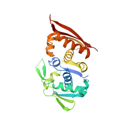
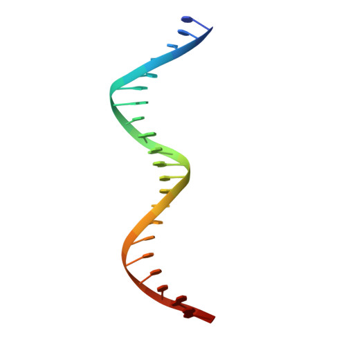
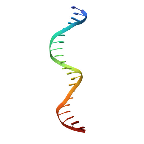
CRYSTAL STRUCTURE OF REPLICATION INITIATOR PROTEIN REPE54 OF MINI-F PLASMID COMPLEXED WITH AN ITERON DNA


Experimental Data Snapshot

  • Method: X-RAY DIFFRACTION
  • Resolution: 2.60 Å
  • R-Value Free: 0.274 
  • R-Value Work: 0.213 

wwPDB Validation   3D Report Full Report


This is version 1.5 of the entry. See complete history


Literature

Crystal structure of a prokaryotic replication initiator protein bound to DNA at 2.6 A resolution.

Komori, H.Matsunaga, F.Higuchi, Y.Ishiai, M.Wada, C.Miki, K.

(1999) EMBO J 18: 4597-4607

  • DOI: https://doi.org/10.1093/emboj/18.17.4597
  • Primary Citation of Related Structures:  
    1REP

  • PubMed Abstract: 

    The initiator protein (RepE) of F factor, a plasmid involved in sexual conjugation in Escherichia coli, has dual functions during the initiation of DNA replication which are determined by whether it exists as a dimer or as a monomer. A RepE monomer functions as a replication initiator, but a RepE dimer functions as an autogenous repressor. We have solved the crystal structure of the RepE monomer bound to an iteron DNA sequence of the replication origin of plasmid F. The RepE monomer consists of topologically similar N- and C-terminal domains related to each other by internal pseudo 2-fold symmetry, despite the lack of amino acid similarities between the domains. Both domains bind to the two major grooves of the iteron (19 bp) with different binding affinities. The C-terminal domain plays the leading role in this binding, while the N-terminal domain has an additional role in RepE dimerization. The structure also suggests that superhelical DNA induced at the origin of plasmid F by four RepEs and one HU dimer has an essential role in the initiation of DNA replication.


  • Organizational Affiliation

    Department of Chemistry, Graduate School of Science, Kyoto University, Sakyo-ku, Kyoto 606-8502, USA.


Macromolecules

Find similar proteins by:  (by identity cutoff)  |  3D Structure
Entity ID: 3
MoleculeChains Sequence LengthOrganismDetailsImage
PROTEIN (REPLICATION INITIATION PROTEIN)251Escherichia coliMutation(s): 1 
UniProt
Find proteins for P03856 (Escherichia coli (strain K12))
Explore P03856 
Go to UniProtKB:  P03856
Entity Groups  
Sequence Clusters30% Identity50% Identity70% Identity90% Identity95% Identity100% Identity
UniProt GroupP03856
Sequence Annotations
Expand
  • Reference Sequence

Find similar nucleic acids by:  Sequence   |   3D Structure  

Entity ID: 1
MoleculeChains LengthOrganismImage
DNA (5'-D(*CP*CP*TP*GP*TP*GP*AP*CP*AP*AP*AP*TP*TP*GP*CP*CP*CP*TP*CP*AP*GP*T)-3')22N/A
Sequence Annotations
Expand
  • Reference Sequence

Find similar nucleic acids by:  Sequence   |   3D Structure  

Entity ID: 2
MoleculeChains LengthOrganismImage
DNA (5'-D(*CP*TP*GP*AP*GP*GP*GP*CP*AP*AP*TP*TP*TP*GP*TP*CP*AP*CP*AP*GP*GP*T)-3')22N/A
Sequence Annotations
Expand
  • Reference Sequence
Small Molecules
Ligands 1 Unique
IDChains Name / Formula / InChI Key2D Diagram3D Interactions
MG
Query on MG

Download Ideal Coordinates CCD File 
D [auth C]MAGNESIUM ION
Mg
JLVVSXFLKOJNIY-UHFFFAOYSA-N
Experimental Data & Validation

Experimental Data

  • Method: X-RAY DIFFRACTION
  • Resolution: 2.60 Å
  • R-Value Free: 0.274 
  • R-Value Work: 0.213 
  • Space Group: C 1 2 1
Unit Cell:
Length ( Å )Angle ( ˚ )
a = 108.435α = 90
b = 81.853β = 121.53
c = 73.891γ = 90
Software Package:
Software NamePurpose
CCP4model building
X-PLORrefinement
DENZOdata reduction
SCALEPACKdata scaling
CCP4phasing

Structure Validation

View Full Validation Report


Entry History 

Deposition Data

Revision History  (Full details and data files)

  • Version 1.0: 2000-02-09
    Type: Initial release
  • Version 1.1: 2008-04-26
    Changes: Version format compliance
  • Version 1.2: 2011-07-13
    Changes: Version format compliance
  • Version 1.3: 2017-11-29
    Changes: Derived calculations
  • Version 1.4: 2018-03-14
    Changes: Database references
  • Version 1.5: 2023-12-27
    Changes: Data collection, Database references, Derived calculations