1RC2 | pdb_00001rc2

2.5 Angstrom Resolution X-ray Structure of Aquaporin Z


Experimental Data Snapshot

  • Method: X-RAY DIFFRACTION
  • Resolution: 2.50 Å
  • R-Value Free: 
    0.268 (Depositor) 
  • R-Value Work: 
    0.227 (Depositor) 

Starting Model: experimental
View more details

wwPDB Validation 3D Report Full Report

Validation slider image for 1RC2

This is version 1.4 of the entry. See complete history

Literature

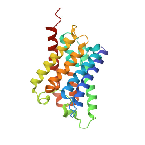
Architecture and selectivity in aquaporins: 2.5 a X-ray structure of aquaporin Z

Savage, D.F.Egea, P.F.Robles-Colmenares, Y.O'Connell III, J.D.Stroud, R.M.

(2003) PLoS Biol 1: 334-340

  • DOI: https://doi.org/10.1371/journal.pbio.0000072
  • Primary Citation Related Structures: 
    1RC2

  • PubMed Abstract: 

    Aquaporins are a family of water and small molecule channels found in organisms ranging from bacteria to animals. One of these channels, the E. coli protein aquaporin Z (AqpZ), has been shown to selectively conduct only water at high rates. We have expressed, purified, crystallized, and solved the X-ray structure of AqpZ. The 2.5 A resolution structure of AqpZ suggests aquaporin selectivity results both from a steric mechanism due to pore size and from specific amino acid substitutions that regulate the preference for a hydrophobic or hydrophilic substrate. This structure provides direct evidence on the molecular mechanisms of specificity between water and glycerol in this family of channels from a single species. It is to our knowledge the first atomic resolution structure of a recombinant aquaporin and so provides a platform for combined genetic, mutational, functional, and structural determinations of the mechanisms of aquaporins and, more generally, the assembly of multimeric membrane proteins.


  • Organizational Affiliation
    • Department of Biochemistry and Biophysics, University of California School of Medicine, San Francisco, California, USA.

Macromolecule Content 

  • Total Structure Weight: 48.6 kDa 
  • Atom Count: 3,530 
  • Modeled Residue Count: 462 
  • Deposited Residue Count: 462 
  • Unique protein chains: 1

Macromolecules

Find similar proteins by:|  3D Structure
Entity ID: 1
MoleculeChains  Sequence LengthOrganismDetailsImage
Aquaporin ZA [auth B],
B [auth A]
231Escherichia coliMutation(s): 0 
Gene Names: AQPZBNIPB0875C1009SF0832S0873
Membrane Entity: Yes 
UniProt
Find proteins for P60844 (Escherichia coli (strain K12))
Explore P60844 
Go to UniProtKB:  P60844
Entity Groups
Sequence Clusters30% Identity50% Identity70% Identity90% Identity95% Identity100% Identity
UniProt GroupP60844
Sequence Annotations
Expand
Reference Sequence

Experimental Data & Validation

Experimental Data

  • Method: X-RAY DIFFRACTION
  • Resolution: 2.50 Å
  • R-Value Free:  0.268 (Depositor) 
  • R-Value Work:  0.227 (Depositor) 
Space Group: P 4
Unit Cell:
Length ( Å )Angle ( ˚ )
a = 93.55α = 90
b = 93.55β = 90
c = 80.38γ = 90
Software Package:
Software NamePurpose
DENZOdata reduction
SCALEPACKdata scaling
CNSrefinement
CNSphasing

Structure Validation

View Full Validation Report


Entry History 

Deposition Data

Revision History  (Full details and data files)

  • Version 1.0: 2003-11-25
    Type: Initial release
  • Version 1.1: 2008-04-29
    Changes: Version format compliance
  • Version 1.2: 2011-07-13
    Changes: Derived calculations, Version format compliance
  • Version 1.3: 2020-07-29
    Type: Remediation
    Reason: Carbohydrate remediation
    Changes: Data collection, Derived calculations, Structure summary
  • Version 1.4: 2023-08-23
    Changes: Data collection, Database references, Refinement description, Structure summary