1R26

Crystal structure of thioredoxin from Trypanosoma brucei brucei


Experimental Data Snapshot

  • Method: X-RAY DIFFRACTION
  • Resolution: 1.40 Å
  • R-Value Free: 0.198 
  • R-Value Work: 0.168 
  • R-Value Observed: 0.169 

Starting Model: experimental
View more details

wwPDB Validation   3D Report Full Report


This is version 1.3 of the entry. See complete history


Literature

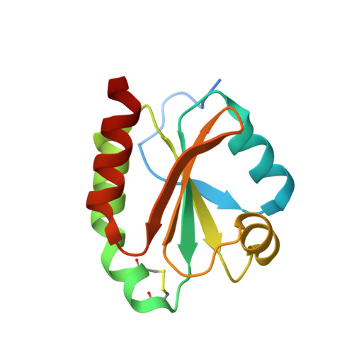
Structure of thioredoxin from Trypanosoma brucei brucei

Friemann, R.Schmidt, H.Ramaswamy, S.Forstner, M.Krauth-Siegel, R.L.Eklund, H.

(2003) FEBS Lett 554: 301-305

  • DOI: https://doi.org/10.1016/s0014-5793(03)01173-6
  • Primary Citation of Related Structures:  
    1R26

  • PubMed Abstract: 

    The three-dimensional structure of thioredoxin from Trypanosoma brucei brucei has been determined at 1.4 A resolution. The overall structure is more similar to that of human thioredoxin than to any other thioredoxin structure. The most striking difference to other thioredoxins is the absence of a buried carboxylate behind the active site cysteines. Instead of the common Asp, there is a Trp that binds an ordered water molecule probably involved in the protonation/deprotonation of the more buried cysteine during catalysis. The conserved Trp in the WCGPC sequence motif has an exposed position that can interact with target proteins.


  • Organizational Affiliation

    Department of Molecular Biosciences, Swedish University of Agricultural Sciences, Biomedical Center, Box 590, S-75124 Uppsala, Sweden.


Macromolecules
Find similar proteins by:  (by identity cutoff)  |  3D Structure
Entity ID: 1
MoleculeChains Sequence LengthOrganismDetailsImage
Thioredoxin125Trypanosoma brucei bruceiMutation(s): 0 
Gene Names: trx
UniProt
Find proteins for Q9NG23 (Trypanosoma brucei brucei)
Explore Q9NG23 
Go to UniProtKB:  Q9NG23
Entity Groups  
Sequence Clusters30% Identity50% Identity70% Identity90% Identity95% Identity100% Identity
UniProt GroupQ9NG23
Sequence Annotations
Expand
  • Reference Sequence
Experimental Data & Validation

Experimental Data

  • Method: X-RAY DIFFRACTION
  • Resolution: 1.40 Å
  • R-Value Free: 0.198 
  • R-Value Work: 0.168 
  • R-Value Observed: 0.169 
  • Space Group: P 21 21 21
Unit Cell:
Length ( Å )Angle ( ˚ )
a = 37.878α = 90
b = 51.725β = 90
c = 57.389γ = 90
Software Package:
Software NamePurpose
d*TREKdata scaling
d*TREKdata reduction
AMoREphasing
REFMACrefinement

Structure Validation

View Full Validation Report


Entry History 

Deposition Data

Revision History  (Full details and data files)

  • Version 1.0: 2003-12-09
    Type: Initial release
  • Version 1.1: 2008-04-29
    Changes: Version format compliance
  • Version 1.2: 2011-07-13
    Changes: Source and taxonomy, Version format compliance
  • Version 1.3: 2023-08-23
    Changes: Data collection, Database references, Refinement description