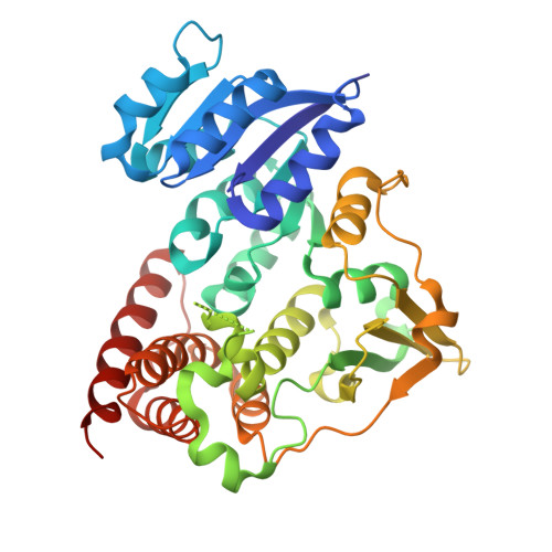
Crystal structure of 1-deoxy-d-xylulose-5-phosphate reductoisomerase from Zymomonas mobilis at 1.9-A resolution.
Ricagno, S., Grolle, S., Bringer-Meyer, S., Sahm, H., Lindqvist, Y., Schneider, G.(2004) Biochim Biophys Acta 1698: 37-44
- PubMed: 15063313
- DOI: https://doi.org/10.1016/j.bbapap.2003.10.006
- Primary Citation of Related Structures:
1R0K, 1R0L - PubMed Abstract:
1-Deoxy-d-xylulose-5-phosphate reductoisomerase (DXR) is the second enzyme in the non-mevalonate pathway of isoprenoid biosynthesis. The structure of the apo-form of this enzyme from Zymomonas mobilis has been solved and refined to 1.9-A resolution, and that of a binary complex with the co-substrate NADPH to 2.7-A resolution. The subunit of DXR consists of three domains. Residues 1-150 form the NADPH binding domain, which is a variant of the typical dinucleotide-binding fold. The second domain comprises a four-stranded mixed beta-sheet, with three helices flanking the sheet. Most of the putative active site residues are located on this domain. The C-terminal domain (residues 300-386) folds into a four-helix bundle. In solution and in the crystal, the enzyme forms a homo-dimer. The interface between the two monomers is formed predominantly by extension of the sheet in the second domain. The adenosine phosphate moiety of NADPH binds to the nucleotide-binding fold in the canonical way. The adenine ring interacts with the loop after beta1 and with the loops between alpha2 and beta2 and alpha5 and beta5. The nicotinamide ring is disordered in crystals of this binary complex. Comparisons to Escherichia coli DXR show that the two enzymes are very similar in structure, and that the active site architecture is highly conserved. However, there are differences in the recognition of the adenine ring of NADPH in the two enzymes.
- Department of Medical Biochemistry and Biophysics, Division of Molecular Structural Biology, Karolinska Institutet, Scheelevagen 2, S-171-77 Stockholm, Sweden.
Organizational Affiliation:


