1QZ9 | pdb_00001qz9

The Three Dimensional Structure of Kynureninase from Pseudomonas fluorescens


Experimental Data Snapshot

  • Method: X-RAY DIFFRACTION
  • Resolution: 1.85 Å
  • R-Value Free: 
    0.192 (Depositor), 0.270 (DCC) 
  • R-Value Work: 
    0.153 (Depositor) 
  • R-Value Observed: 
    0.155 (Depositor) 

Starting Model: experimental
View more details

wwPDB Validation 3D Report Full Report

Validation slider image for 1QZ9

Ligand Structure Quality Assessment 


This is version 1.3 of the entry. See complete history

Literature

Three-Dimensional Structure of Kynureninase from Pseudomonas fluorescens.

Momany, C.Levdikov, V.Blagova, L.Lima, S.Phillips, R.S.

(2004) Biochemistry 43: 1193-1203

  • DOI: https://doi.org/10.1021/bi035744e
  • Primary Citation Related Structures: 
    1QZ9

  • PubMed Abstract: 

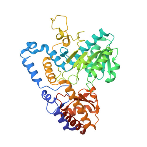
    Kynureninase [E.C. 3.7.1.3] is a pyridoxal-5'-phosphate (PLP)-dependent enzyme that catalyzes the hydrolytic cleavage of l-kynurenine to anthranilic acid and l-alanine. Sequence alignment with other PLP-dependent enzymes indicated that kynureninase is in subgroup IVa of the aminotransferases, along with nifS, CsdB, and serine-pyruvate aminotransferase, which suggests that kynureninase has an aminotransferase fold. Crystals of Pseudomonas fluorescens kynureninase were obtained, and the structure was solved by molecular replacement using the CsdB coordinates combined with multiple isomorphous heavy atom replacement. The coordinates were deposited in the PDB (ID code 1QZ9). The structure, refined to an R factor of 15.5% to 1.85 A resolution, is dimeric and has the aminotransferase fold. The structure also confirms the prediction from sequence alignment that Lys-227 is the PLP-binding residue in P. fluorescens kynureninase. The conserved Asp-201, expected for an aminotransferase fold, is located near the PLP nitrogen, but Asp-132 is also strictly conserved and at a similar distance from the pyridinium nitrogen. Mutagenesis of both conserved aspartic acids shows that both contribute equally to PLP binding, but Asp-201 has a greater role in catalysis. The structure shows that Tyr-226 donates a hydrogen bond to the phosphate of PLP. Unusual among PLP-dependent enzymes, Trp-256, which is also strictly conserved in kynureninases from bacteria to humans, donates a hydrogen bond to the phosphate through the indole N1-hydrogen.


  • Organizational Affiliation
    • Department of Chemistry, University of Georgia, Athens, Georgia 30602, USA.

Macromolecule Content 

  • Total Structure Weight: 46.48 kDa 
  • Atom Count: 3,620 
  • Modeled Residue Count: 404 
  • Deposited Residue Count: 416 
  • Unique protein chains: 1

Macromolecules

Find similar proteins by:|  3D Structure
Entity ID: 1
MoleculeChains  Sequence LengthOrganismDetailsImage
KYNURENINASE416Pseudomonas fluorescensMutation(s): 0 
EC: 3.7.1.3
UniProt
Find proteins for P83788 (Pseudomonas fluorescens)
Explore P83788 
Go to UniProtKB:  P83788
Entity Groups
Sequence Clusters30% Identity50% Identity70% Identity90% Identity95% Identity100% Identity
UniProt GroupP83788
Sequence Annotations
Expand
Reference Sequence

Small Molecules

Ligands 3 Unique
IDChains Name / Formula / InChI Key2D Diagram3D Interactions
P3G

Query on P3G


Download:Ideal Coordinates CCD File
D [auth A]3,6,9,12,15-PENTAOXAHEPTADECANE
C12 H26 O5
HYDWALOBQJFOMS-UHFFFAOYSA-N
PLP

Query on PLP


Download:Ideal Coordinates CCD File
C [auth A]PYRIDOXAL-5'-PHOSPHATE
C8 H10 N O6 P
NGVDGCNFYWLIFO-UHFFFAOYSA-N
CL

Query on CL


Download:Ideal Coordinates CCD File
B [auth A]CHLORIDE ION
Cl
VEXZGXHMUGYJMC-UHFFFAOYSA-M

Experimental Data & Validation

Experimental Data

  • Method: X-RAY DIFFRACTION
  • Resolution: 1.85 Å
  • R-Value Free:  0.192 (Depositor), 0.270 (DCC) 
  • R-Value Work:  0.153 (Depositor) 
  • R-Value Observed: 0.155 (Depositor) 
Space Group: P 31 2 1
Unit Cell:
Length ( Å )Angle ( ˚ )
a = 68.076α = 90
b = 68.076β = 90
c = 137.976γ = 120
Software Package:
Software NamePurpose
REFMACrefinement
DENZOdata reduction
SCALEPACKdata scaling
SOLVEphasing

Structure Validation

View Full Validation Report


Ligand Structure Quality Assessment 


Entry History 

Deposition Data

Revision History  (Full details and data files)

  • Version 1.0: 2004-01-20
    Type: Initial release
  • Version 1.1: 2008-04-29
    Changes: Version format compliance
  • Version 1.2: 2011-07-13
    Changes: Advisory, Derived calculations, Version format compliance
  • Version 1.3: 2023-08-23
    Changes: Data collection, Database references, Derived calculations, Refinement description