1QXA | pdb_00001qxa

Crystal structure of Sortase B complexed with Gly3


Experimental Data Snapshot

  • Method: X-RAY DIFFRACTION
  • Resolution: 2.50 Å
  • R-Value Free: 
    0.235 (Depositor), 0.236 (DCC) 
  • R-Value Work: 
    0.221 (Depositor), 0.221 (DCC) 
  • R-Value Observed: 
    0.221 (Depositor) 

wwPDB Validation 3D Report Full Report

Validation slider image for 1QXA

This is version 1.4 of the entry. See complete history

Literature

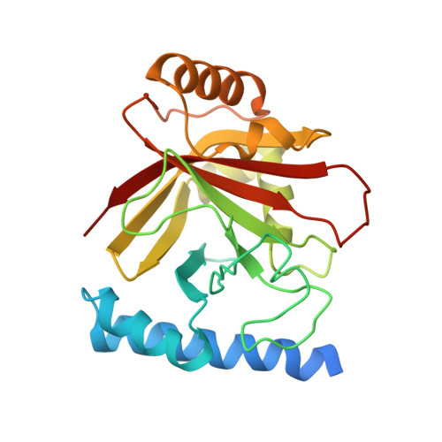
The structure of sortase B, a cysteine transpeptidase that tethers surface protein to the Staphylococcus aureus cell wall

Zong, Y.Mazmanian, S.K.Schneewind, O.Narayana, S.V.

(2004) Structure 12: 105-112

  • DOI: https://doi.org/10.1016/j.str.2003.11.021
  • Primary Citation Related Structures: 
    1QWZ, 1QX6, 1QXA

  • PubMed Abstract: 

    Many surface proteins of Gram-positive bacteria, which play important roles during the pathogenesis of human infections, are anchored to the cell wall envelope by a mechanism requiring sortases. Sortase B, a cysteine transpeptidase from Staphylococcus aureus, cleaves the C-terminal sorting signal of IsdC at the NPQTN motif and tethers the polypeptide to the pentaglycine cell wall cross-bridge. During catalysis, the active site cysteine of sortase and the cleaved substrate form an acyl intermediate, which is then resolved by the amino group of pentaglycine cross-bridges. We report here the crystal structures of SrtBDeltaN30 in complex with two active site inhibitors, MTSET and E64, and with the cell wall substrate analog tripleglycine. These structures reveal, for the first time, the active site disposition and the unique Cys-Arg catalytic machinery of the cysteine transpeptidase, and they also provide useful information for the future design of anti-infective agents against sortases.


  • Organizational Affiliation
    • Center for Biophysical Sciences and Engineering, School of Optometry, University of Alabama at Birmingham, Birmingham, AL 35294, USA.

Macromolecule Content 

  • Total Structure Weight: 28.23 kDa 
  • Atom Count: 1,843 
  • Modeled Residue Count: 217 
  • Deposited Residue Count: 238 
  • Unique protein chains: 2

Macromolecules

Find similar proteins by:|  3D Structure
Entity ID: 1
MoleculeChains  Sequence LengthOrganismDetailsImage
NPQTN specific sortase B235Staphylococcus aureusMutation(s): 0 
EC: 3.4.22.71
UniProt
Find proteins for Q2FZE3 (Staphylococcus aureus (strain NCTC 8325 / PS 47))
Explore Q2FZE3 
Go to UniProtKB:  Q2FZE3
Entity Groups
Sequence Clusters30% Identity50% Identity70% Identity90% Identity95% Identity100% Identity
UniProt GroupQ2FZE3
Sequence Annotations
Expand
Reference Sequence
Find similar proteins by:  Sequence   |   3D Structure  
Entity ID: 2
MoleculeChains  Sequence LengthOrganismDetailsImage
peptide GLY-GLY-GLY3N/AMutation(s): 0 

Small Molecules

Ligands 1 Unique
IDChains Name / Formula / InChI Key2D Diagram3D Interactions
ETM

Query on ETM


Download:Ideal Coordinates CCD File
C [auth A]2-(TRIMETHYLAMMONIUM)ETHYL THIOL
C5 H14 N S
VFUGTBZQGUVGEX-UHFFFAOYSA-O

Experimental Data & Validation

Experimental Data

  • Method: X-RAY DIFFRACTION
  • Resolution: 2.50 Å
  • R-Value Free:  0.235 (Depositor), 0.236 (DCC) 
  • R-Value Work:  0.221 (Depositor), 0.221 (DCC) 
  • R-Value Observed: 0.221 (Depositor) 
Space Group: C 2 2 21
Unit Cell:
Length ( Å )Angle ( ˚ )
a = 57.69α = 90
b = 67.23β = 90
c = 102.58γ = 90
Software Package:
Software NamePurpose
CNSrefinement
SCALEPACKdata scaling
CNSphasing

Structure Validation

View Full Validation Report


Entry History 

Deposition Data

Revision History  (Full details and data files)

  • Version 1.0: 2004-04-06
    Type: Initial release
  • Version 1.1: 2008-04-29
    Changes: Version format compliance
  • Version 1.2: 2011-07-13
    Changes: Version format compliance
  • Version 1.3: 2017-10-11
    Changes: Refinement description
  • Version 1.4: 2024-10-16
    Changes: Data collection, Database references, Derived calculations, Structure summary