1QVS | pdb_00001qvs

Crystal Structure of Haemophilus influenzae H9A mutant Holo Ferric ion-Binding Protein A


Experimental Data Snapshot

  • Method: X-RAY DIFFRACTION
  • Resolution: 2.10 Å
  • R-Value Free: 
    0.226 (Depositor), 0.238 (DCC) 
  • R-Value Work: 
    0.168 (Depositor), 0.180 (DCC) 
  • R-Value Observed: 
    0.171 (Depositor) 

Starting Model: experimental
View more details

wwPDB Validation 3D Report Full Report

Validation slider image for 1QVS

This is version 1.4 of the entry. See complete history

Literature

Presence of ferric hydroxide clusters in mutants of Haemophilus influenzae ferric ion-binding protein A

Shouldice, S.R.Skene, R.J.Dougan, D.R.McRee, D.E.Tari, L.W.Schryvers, A.B.

(2003) Biochemistry 42: 11908-11914

  • DOI: https://doi.org/10.1021/bi035389s
  • Primary Citation Related Structures: 
    1QVS, 1QW0

  • PubMed Abstract: 

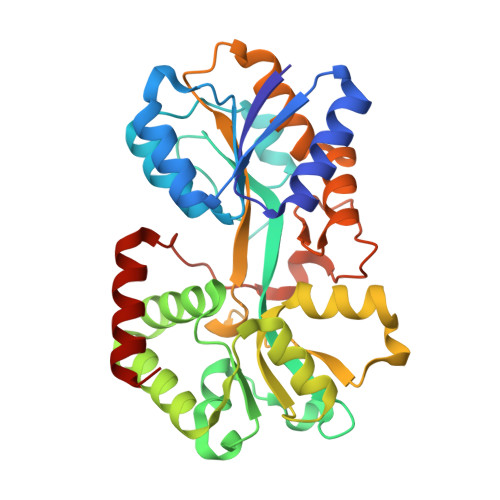
    The periplasmic iron binding protein plays an essential role in the iron uptake pathway of Gram-negative pathogenic bacteria from the Pasteurellaceae and Neisseriaceae families and is critical for survival of these pathogens within the host. In this study, we report the crystal structures of two mutant forms of ferric ion-binding protein A (FbpA) from Haemophilus influenzae with bound multinuclear oxo-metal clusters. Crystals of site-directed mutants in the metal or anion binding ligands contain protein in the open conformation, and two mutant FbpAs, H9A and N175L, contain different cluster arrangements in the iron-binding pocket. The iron clusters are anchored by binding to the two tyrosine ligands (Tyr195 and Tyr196) positioned at the vertex of the iron-binding pocket but are not coordinated by the other metal binding ligands. Our results suggest that the metal clusters may have formed in situ, suggesting that the mutant FbpAs may serve as a simple model for protein-mediated mineralization.


  • Organizational Affiliation
    • Department of Microbiology and Infectious Diseases, University of Calgary, Calgary, Alberta, Canada T2N 4N1.

Macromolecule Content 

  • Total Structure Weight: 34.06 kDa 
  • Atom Count: 2,588 
  • Modeled Residue Count: 308 
  • Deposited Residue Count: 309 
  • Unique protein chains: 1

Macromolecules

Find similar proteins by:|  3D Structure
Entity ID: 1
MoleculeChains  Sequence LengthOrganismDetailsImage
Iron-utilization periplasmic protein309Haemophilus influenzaeMutation(s): 1 
Gene Names: FBPAHITAFBP or HI0097
UniProt
Find proteins for P35755 (Haemophilus influenzae (strain ATCC 51907 / DSM 11121 / KW20 / Rd))
Explore P35755 
Go to UniProtKB:  P35755
Entity Groups
Sequence Clusters30% Identity50% Identity70% Identity90% Identity95% Identity100% Identity
UniProt GroupP35755
Sequence Annotations
Expand
Reference Sequence

Experimental Data & Validation

Experimental Data

  • Method: X-RAY DIFFRACTION
  • Resolution: 2.10 Å
  • R-Value Free:  0.226 (Depositor), 0.238 (DCC) 
  • R-Value Work:  0.168 (Depositor), 0.180 (DCC) 
  • R-Value Observed: 0.171 (Depositor) 
Space Group: P 21 21 2
Unit Cell:
Length ( Å )Angle ( ˚ )
a = 105.999α = 90
b = 75.656β = 90
c = 33.774γ = 90
Software Package:
Software NamePurpose
REFMACrefinement
HKL-2000data reduction
CCP4data scaling
MOLREPphasing

Structure Validation

View Full Validation Report


Entry History 

Deposition Data

Revision History  (Full details and data files)

  • Version 1.0: 2003-11-04
    Type: Initial release
  • Version 1.1: 2008-04-29
    Changes: Version format compliance
  • Version 1.2: 2011-07-13
    Changes: Version format compliance
  • Version 1.3: 2021-10-27
    Changes: Database references, Derived calculations
  • Version 1.4: 2023-08-16
    Changes: Data collection, Refinement description