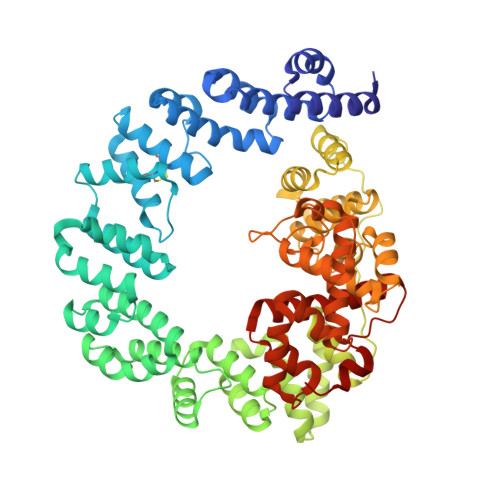
1QTE

CRYSTAL STRUCTURE OF THE 70 KDA SOLUBLE LYTIC TRANSGLYCOSYLASE SLT70 FROM ESCHERICHIA COLI AT 1.90 A RESOLUTION IN COMPLEX WITH A 1,6-ANHYDROMUROTRIPEPTIDE


Experimental Data Snapshot

  • Method: X-RAY DIFFRACTION
  • Resolution: 1.90 Å
  • R-Value Free: 0.197 
  • R-Value Work: 0.171 
  • R-Value Observed: 0.171 

wwPDB Validation   3D Report Full Report


Ligand Structure Quality Assessment 


This is version 1.4 of the entry. See complete history


Literature

High resolution crystal structures of the Escherichia coli lytic transglycosylase Slt70 and its complex with a peptidoglycan fragment.

van Asselt, E.J.Thunnissen, A.M.Dijkstra, B.W.

(1999) J Mol Biol 291: 877-898

  • DOI: https://doi.org/10.1006/jmbi.1999.3013
  • Primary Citation of Related Structures:  
    1QSA, 1QTE

  • PubMed Abstract: 

    The 70 kDa soluble lytic transglycosylase (Slt70) from Escherichia coli is an exo-muramidase, that catalyses the cleavage of the glycosidic bonds between N -acetylmuramic acid and N -acetylglucosamine residues in peptidoglycan, the main structural component of the bacterial cell wall. This cleavage is accompanied by the formation of a 1,6-anhydro bond between the C1 and O6 atoms in the N -acetylmuramic acid residue (anhMurNAc). Crystallographic studies at medium resolution revealed that Slt70 is a multi-domain protein consisting of a large ring-shaped alpha-superhelix with on top a catalytic domain, which resembles the fold of goose-type lysozyme. Here we report the crystal structures of native Slt70 and of its complex with a 1,6-anhydromuropeptide solved at nominal resolutions of 1.65 A and 1.90 A, respectively. The high resolution native structure reveals the details on the hydrogen bonds, electrostatic and hydrophobic interactions that stabilise the catalytic domain and the alpha-superhelix. The building-block of the alpha-superhelix is an "up-down-up-down" four-alpha-helix bundle involving both parallel and antiparallel helix pairs. Stabilisation of the fold is provided through an extensive packing of apolar atoms, mostly from leucine and alanine residues. It lacks, however, an internal consensus sequence that characterises other super-secondary helical folds like the beta-helix in pectate lyase or the (beta-alpha)-helix in the ribonuclease inhibitor. The 1, 6-anhydromuropeptide product binds in a shallow groove adjacent to the peptidoglycan-binding groove of the catalytic domain. The groove is formed by conserved residues at the interface of the catalytic domain and the alpha-superhelix. The structure of the Slt70-1, 6-anhydromuropeptide complex confirms the presence of a specific binding-site for the peptide moieties of the peptidoglycan and it substantiates the notion that Slt70 starts the cleavage reaction at the anhMurNAc end of the peptidoglycan.


  • Organizational Affiliation

    University of Groningen, Nijenborgh 4, Groningen, 9747 AG, The Netherlands.


Macromolecules
Find similar proteins by:  (by identity cutoff)  |  3D Structure
Entity ID: 1
MoleculeChains Sequence LengthOrganismDetailsImage
SOLUBLE LYTIC TRANSGLYCOSYLASE SLT70618Escherichia coliMutation(s): 0 
EC: 3.2.1
UniProt
Find proteins for P0AGC3 (Escherichia coli (strain K12))
Explore P0AGC3 
Go to UniProtKB:  P0AGC3
Entity Groups  
Sequence Clusters30% Identity50% Identity70% Identity90% Identity95% Identity100% Identity
UniProt GroupP0AGC3
Sequence Annotations
Expand
  • Reference Sequence
Small Molecules
Ligands 7 Unique
IDChains Name / Formula / InChI Key2D Diagram3D Interactions
AH0
Query on AH0

Download Ideal Coordinates CCD File 
R [auth A]2-(2-ACETYLAMINO-4-HYDROXY-6,8-DIOXA-BICYCLO[3.2.1]OCT-3-YLOXY)-PROPIONIC ACID
C11 H17 N O7
ZFEGYUMHFZOYIY-YVNCZSHWSA-N
NAG
Query on NAG

Download Ideal Coordinates CCD File 
B [auth A]2-acetamido-2-deoxy-beta-D-glucopyranose
C8 H15 N O6
OVRNDRQMDRJTHS-FMDGEEDCSA-N
SO4
Query on SO4

Download Ideal Coordinates CCD File 
C [auth A]
D [auth A]
E [auth A]
F [auth A]
G [auth A]
C [auth A],
D [auth A],
E [auth A],
F [auth A],
G [auth A],
H [auth A],
I [auth A],
J [auth A],
K [auth A],
L [auth A],
M [auth A],
N [auth A]
SULFATE ION
O4 S
QAOWNCQODCNURD-UHFFFAOYSA-L
GOL
Query on GOL

Download Ideal Coordinates CCD File 
S [auth A],
T [auth A],
U [auth A],
V [auth A],
W [auth A]
GLYCEROL
C3 H8 O3
PEDCQBHIVMGVHV-UHFFFAOYSA-N
ALA
Query on ALA

Download Ideal Coordinates CCD File 
P [auth A]ALANINE
C3 H7 N O2
QNAYBMKLOCPYGJ-REOHCLBHSA-N
DAL
Query on DAL

Download Ideal Coordinates CCD File 
Q [auth A]D-ALANINE
C3 H7 N O2
QNAYBMKLOCPYGJ-UWTATZPHSA-N
ACT
Query on ACT

Download Ideal Coordinates CCD File 
O [auth A]ACETATE ION
C2 H3 O2
QTBSBXVTEAMEQO-UHFFFAOYSA-M
Experimental Data & Validation

Experimental Data

  • Method: X-RAY DIFFRACTION
  • Resolution: 1.90 Å
  • R-Value Free: 0.197 
  • R-Value Work: 0.171 
  • R-Value Observed: 0.171 
  • Space Group: P 21 21 21
Unit Cell:
Length ( Å )Angle ( ˚ )
a = 78.266α = 90
b = 87.514β = 90
c = 132.947γ = 90
Software Package:
Software NamePurpose
DENZOdata reduction
SCALEPACKdata scaling
X-PLORmodel building
X-PLORrefinement
X-PLORphasing

Structure Validation

View Full Validation Report


Ligand Structure Quality Assessment 


Entry History 

Deposition Data

Revision History  (Full details and data files)

  • Version 1.0: 1999-08-31
    Type: Initial release
  • Version 1.1: 2008-04-27
    Changes: Version format compliance
  • Version 1.2: 2011-07-13
    Changes: Non-polymer description, Version format compliance
  • Version 1.3: 2020-07-29
    Type: Remediation
    Reason: Carbohydrate remediation
    Changes: Data collection, Derived calculations, Structure summary
  • Version 1.4: 2024-10-16
    Changes: Data collection, Database references, Structure summary