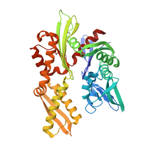
1QQN

D206S MUTANT OF BOVINE 70 KILODALTON HEAT SHOCK PROTEIN


Experimental Data Snapshot

  • Method: X-RAY DIFFRACTION
  • Resolution: 1.90 Å
  • R-Value Free: 0.229 
  • R-Value Work: 0.197 
  • R-Value Observed: 0.197 

wwPDB Validation   3D Report Full Report


This is version 1.6 of the entry. See complete history


Literature

Mapping the role of active site residues for transducing an ATP-induced conformational change in the bovine 70-kDa heat shock cognate protein.

Johnson, E.R.McKay, D.B.

(1999) Biochemistry 38: 10823-18830

  • DOI: https://doi.org/10.1021/bi990816g
  • Primary Citation of Related Structures:  
    1QQM, 1QQN, 1QQO

  • PubMed Abstract: 

    ATP binding induces a conformational change in 70-kDa heat shock proteins (Hsp70s) that facilitates release of bound polypeptides. Using the bovine heat shock cognate protein (Hsc70) as a representative of the Hsp70 family, we have characterized the effect of mutations on the coupling between ATP binding and the nucleotide-induced conformational change. Steady-state solution small-angle X-ray scattering and kinetic fluorescence measurements on a 60-kDa fragment of Hsc70 show that point mutations K71M, E175S, D199S, and D206S in the nucleotide binding cleft impair the ability of ATP to induce a conformational change. A secondary mutation in the peptide binding domain, E543K, "rescues" the ATP-induced transition for three of these mutations (E175S/E543K, D199S/E543K, and D206S/E543K) but not for K71M/E543K. Analysis of kinetics of the ATPase cycle confirm that these effects do not result from unexpectedly rapid ATP hydrolysis or slow ATP binding. Crystallographic structures of E175S, D199S, and D206S mutant ATPase fragment proteins show that the mutations do not perturb the tertiary structure of the protein but do significantly alter the protein-ligand interactions, due in part to an apparent charge compensation effect whereby mutating a (probably) negatively charged carboxyl group to a neutral serine displaces a K+ ion from the nucleotide binding cleft in two out of three cases (E175S and D199S but not D206S).


  • Organizational Affiliation

    Department of Structural Biology, Stanford University School of Medicine, California 94305-5126, USA.


Macromolecules
Find similar proteins by:  (by identity cutoff)  |  3D Structure
Entity ID: 1
MoleculeChains Sequence LengthOrganismDetailsImage
D206S MUTANT OF BOVINE 70 KILODALTON HEAT SHOCK PROTEIN378Bos taurusMutation(s): 1 
EC: 3.6.4.10
UniProt
Find proteins for P19120 (Bos taurus)
Explore P19120 
Go to UniProtKB:  P19120
Entity Groups  
Sequence Clusters30% Identity50% Identity70% Identity90% Identity95% Identity100% Identity
UniProt GroupP19120
Sequence Annotations
Expand
  • Reference Sequence
Small Molecules
Ligands 5 Unique
IDChains Name / Formula / InChI Key2D Diagram3D Interactions
ADP
Query on ADP

Download Ideal Coordinates CCD File 
H [auth A]ADENOSINE-5'-DIPHOSPHATE
C10 H15 N5 O10 P2
XTWYTFMLZFPYCI-KQYNXXCUSA-N
PO4
Query on PO4

Download Ideal Coordinates CCD File 
G [auth A]PHOSPHATE ION
O4 P
NBIIXXVUZAFLBC-UHFFFAOYSA-K
K
Query on K

Download Ideal Coordinates CCD File 
B [auth A],
C [auth A]
POTASSIUM ION
K
NPYPAHLBTDXSSS-UHFFFAOYSA-N
CL
Query on CL

Download Ideal Coordinates CCD File 
D [auth A],
E [auth A]
CHLORIDE ION
Cl
VEXZGXHMUGYJMC-UHFFFAOYSA-M
MG
Query on MG

Download Ideal Coordinates CCD File 
F [auth A]MAGNESIUM ION
Mg
JLVVSXFLKOJNIY-UHFFFAOYSA-N
Binding Affinity Annotations 
IDSourceBinding Affinity
ADP BindingDB:  1QQN Kd: min: 320, max: 324 (nM) from 2 assay(s)
IC50: 280 (nM) from 1 assay(s)
Experimental Data & Validation

Experimental Data

  • Method: X-RAY DIFFRACTION
  • Resolution: 1.90 Å
  • R-Value Free: 0.229 
  • R-Value Work: 0.197 
  • R-Value Observed: 0.197 
  • Space Group: P 21 21 21
Unit Cell:
Length ( Å )Angle ( ˚ )
a = 143.768α = 90
b = 64.424β = 90
c = 46.486γ = 90
Software Package:
Software NamePurpose
CNSrefinement
SCALEPACKdata scaling
CNSphasing

Structure Validation

View Full Validation Report


Entry History 

Deposition Data

Revision History  (Full details and data files)

  • Version 1.0: 1999-09-15
    Type: Initial release
  • Version 1.1: 2008-04-27
    Changes: Version format compliance
  • Version 1.2: 2011-07-13
    Changes: Version format compliance
  • Version 1.3: 2017-10-04
    Changes: Refinement description
  • Version 1.4: 2018-01-31
    Changes: Experimental preparation
  • Version 1.5: 2018-03-14
    Changes: Database references
  • Version 1.6: 2024-02-14
    Changes: Data collection, Database references, Derived calculations