1QLL | pdb_00001qll

Piratoxin-II (Prtx-II) - a K49 PLA2 from Bothrops pirajai


Experimental Data Snapshot

  • Method: X-RAY DIFFRACTION
  • Resolution: 2.04 Å
  • R-Value Free: 
    0.268 (Depositor) 
  • R-Value Work: 
    0.176 (Depositor), 0.172 (DCC) 

Starting Model: experimental
View more details

wwPDB Validation 3D Report Full Report

Validation slider image for 1QLL

Ligand Structure Quality Assessment 


This is version 1.5 of the entry. See complete history

Literature

Structural Basis for Low Catalytic Activity in Lys49 Phospholipases A2-A Hypothesis: The Crystal Structure of Piratoxin II Complexed to Fatty Acid

Lee, W.H.Da Silva Giotto, M.T.Marangoni, S.Toyama, M.H.Polikarpov, I.Garratt, R.C.

(2001) Biochemistry 40: 28

  • DOI: https://doi.org/10.1021/bi0010470
  • Primary Citation Related Structures: 
    1QLL

  • PubMed Abstract: 

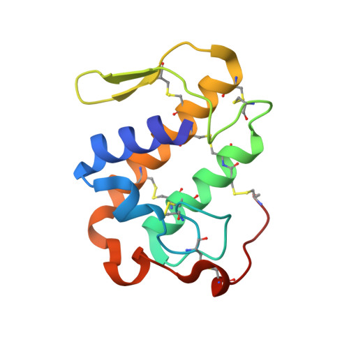
    Asp49 plays a fundamental role in supporting catalysis by phospholipases A2 by coordinating the calcium ion which aids in the stabilization of the tetrahedral intermediate. In several myotoxins from the venoms of Viperidae snakes, this aspartic acid is substituted by lysine. The loss of calcium binding capacity by these mutants has become regarded as the standard explanation for their greatly reduced or nonexistent phospholipolytic activity. Here we describe the crystal structure of one such Lys49 PLA2, piratoxin-II, in which a fatty acid molecule is observed within the substrate channel. This suggests that such toxins may be active enzymes in which catalysis is interrupted at the stage of substrate release. Comparison of the present structure with other PLA2s, both active and inactive, identifies Lys122 as one of the likely causes of the increased affinity for fatty acid in Lys49 enzymes. Its interaction with the mainchain carbonyl of Cys29 is expected to lead to hyperpolarization of the peptide bond between residues 29 and 30 leading to an increased affinity for the fatty acid headgroup. This strongly bound fatty acid may serve as an anchor to secure the toxin within the membrane thus facilitating its pathological effects.


  • Organizational Affiliation
    • Laboratório Nacional de Luz Síncrotron, Caixa Postal 6192, 13083-970 Campinas, São Paulo, Brazil.

Macromolecule Content 

  • Total Structure Weight: 28 kDa 
  • Atom Count: 2,240 
  • Modeled Residue Count: 242 
  • Deposited Residue Count: 242 
  • Unique protein chains: 1

Macromolecules

Find similar proteins by:|  3D Structure
Entity ID: 1
MoleculeChains  Sequence LengthOrganismDetailsImage
PHOSPHOLIPASE A2
A, B
121Bothrops pirajaiMutation(s): 0 
EC: 3.1.1.4
UniProt
Find proteins for P82287 (Bothrops pirajai)
Explore P82287 
Go to UniProtKB:  P82287
Entity Groups
Sequence Clusters30% Identity50% Identity70% Identity90% Identity95% Identity100% Identity
UniProt GroupP82287
Sequence Annotations
Expand
Reference Sequence

Experimental Data & Validation

Experimental Data

  • Method: X-RAY DIFFRACTION
  • Resolution: 2.04 Å
  • R-Value Free:  0.268 (Depositor) 
  • R-Value Work:  0.176 (Depositor), 0.172 (DCC) 
Space Group: P 1 21 1
Unit Cell:
Length ( Å )Angle ( ˚ )
a = 46.19α = 90
b = 60.36β = 96.05
c = 58.74γ = 90
Software Package:
Software NamePurpose
REFMACrefinement
DENZOdata reduction
SCALEPACKdata scaling
AMoREphasing

Structure Validation

View Full Validation Report


Ligand Structure Quality Assessment 


Entry History 

Deposition Data

Revision History  (Full details and data files)

  • Version 1.0: 2000-09-07
    Type: Initial release
  • Version 1.1: 2011-05-08
    Changes: Version format compliance
  • Version 1.2: 2011-07-13
    Changes: Version format compliance
  • Version 1.3: 2019-03-06
    Changes: Data collection, Experimental preparation, Other
  • Version 1.4: 2023-12-13
    Changes: Data collection, Database references, Derived calculations, Other, Refinement description
  • Version 1.5: 2024-11-13
    Changes: Structure summary