1QKE | pdb_00001qke

ERABUTOXIN


Experimental Data Snapshot

  • Method: X-RAY DIFFRACTION
  • Resolution: 1.50 Å
  • R-Value Work: 
    0.162 (DCC) 

Starting Model: experimental
View more details

wwPDB Validation 3D Report Full Report

Validation slider image for 1QKE

This is version 1.5 of the entry. See complete history

Literature

Structure of dimeric and monomeric erabutoxin a refined at 1.5 A resolution.

Nastopoulos, V.Kanellopoulos, P.N.Tsernoglou, D.

(1998) Acta Crystallogr D Biol Crystallogr 54: 964-974

  • DOI: https://doi.org/10.1107/s0907444998005125
  • Primary Citation Related Structures: 
    1QKD, 1QKE

  • PubMed Abstract: 

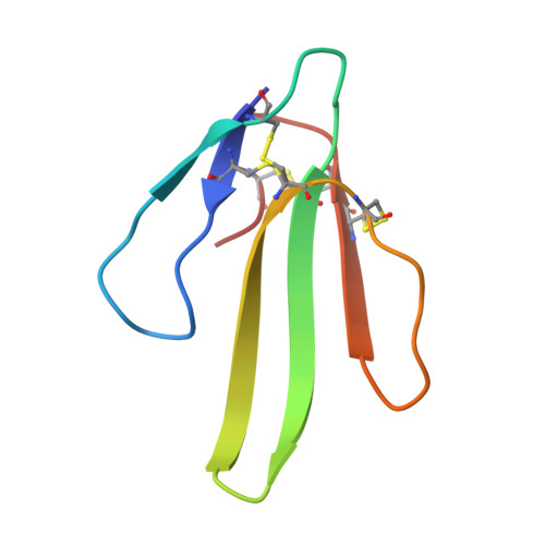
    Erabutoxin a has been crystallized in its monomeric and dimeric forms. The structures were refined at 1.50 and 1.49 A resolution, respectively, using synchrotron radiation data. The crystals belong to space group P212121, with cell dimensions a = 49.84, b = 46.62, c = 21.22 A for the monomer and a = 55.32, b = 53.54, c = 40.76 A for the dimer. Using starting models from earlier structure determinations, the monomeric structure refined to an R value of 16.7% (8004 unique reflections, 17.0-1.50 A resolution range), while the dimeric structure has been solved by the molecular-replacement method with a final R value of 16.9% (19 444 unique reflections, 17.4-1.49 A resolution range). The high-resolution electron-density maps clearly revealed significant discrete disorder in the proteins and allowed an accurate determination of the solvent structure. For the monomer, the side chains of six residues were modelled with alternate conformers and 106 sites for water molecules and one site for a sulfate ion were included in the final model, whereas for the dimer, 206 sites for water molecules were included and both C-terminal residues together with the side chains of 11 residues adopted alternative conformations. A comparison was made with earlier structure determinations. The features of the solvent structure of the erabutoxin molecules are discussed in detail.


  • Organizational Affiliation
    • Department of Chemistry, University of Patras, 26500 Patras, Greece. nastopoulos@upatras.gr

Macromolecule Content 

  • Total Structure Weight: 6.95 kDa 
  • Atom Count: 600 
  • Modeled Residue Count: 62 
  • Deposited Residue Count: 62 
  • Unique protein chains: 1

Macromolecules

Find similar proteins by:|  3D Structure
Entity ID: 1
MoleculeChains  Sequence LengthOrganismDetailsImage
ERABUTOXIN A62Laticauda semifasciataMutation(s): 0 
UniProt
Find proteins for P60775 (Laticauda semifasciata)
Explore P60775 
Go to UniProtKB:  P60775
Entity Groups
Sequence Clusters30% Identity50% Identity70% Identity90% Identity95% Identity100% Identity
UniProt GroupP60775
Sequence Annotations
Expand
Reference Sequence

Small Molecules

Ligands 1 Unique
IDChains Name / Formula / InChI Key2D Diagram3D Interactions
SO4

Query on SO4


Download:Ideal Coordinates CCD File
B [auth A]SULFATE ION
O4 S
QAOWNCQODCNURD-UHFFFAOYSA-L

Experimental Data & Validation

Experimental Data

  • Method: X-RAY DIFFRACTION
  • Resolution: 1.50 Å
  • R-Value Work:  0.162 (DCC) 
Space Group: P 21 21 21
Unit Cell:
Length ( Å )Angle ( ˚ )
a = 49.84α = 90
b = 46.62β = 90
c = 21.22γ = 90
Software Package:
Software NamePurpose
DENZOdata reduction
SCALEPACKdata scaling
X-PLORmodel building
TNTrefinement
X-PLORrefinement
X-PLORphasing

Structure Validation

View Full Validation Report


Entry History 

Deposition Data

Revision History  (Full details and data files)

  • Version 1.0: 1999-02-16
    Type: Initial release
  • Version 1.1: 2008-03-24
    Changes: Version format compliance
  • Version 1.2: 2011-07-13
    Changes: Version format compliance
  • Version 1.3: 2016-10-26
    Changes: Other
  • Version 1.4: 2023-08-09
    Changes: Data collection, Database references, Derived calculations, Refinement description
  • Version 1.5: 2024-10-23
    Changes: Data collection, Structure summary