Structure, dynamics and hydration of the nogalamycin-d(ATGCAT)2Complex determined by NMR and molecular dynamics simulations in solution.
Williams, H.E., Searle, M.S.(1999) J Mol Biology 290: 699-716
- PubMed: 10395824 Search on PubMed
- DOI: https://doi.org/10.1006/jmbi.1999.2903
- Primary Citation Related Structures:
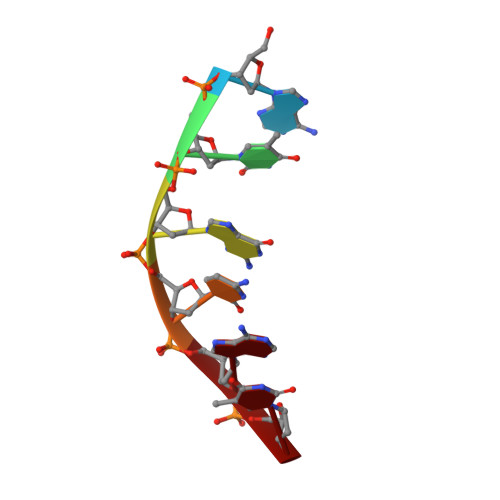
1QCH - PubMed Abstract:
The structure of the 1:1 nogalamycin:d(ATGCAT)2 complex has been determined in solution from high-resolution NMR data and restrained molecular dynamics (rMD) simulations using an explicit solvation model. The antibiotic intercalates at the 5'-TpG step with the nogalose lying along the minor groove towards the centre of the duplex. Many drug-DNA nuclear Overhauser enhancements (NOEs) in the minor groove are indicative of hydrophobic interactions over the TGCA sequence. Steric occlusion prevents a second nogalamycin molecule from binding at the symmetry-related 5'-CpA site, leading to the conclusion that the observed binding orientation in this complex is the preferred orientation free of the complication of end-effects (drug molecules occupy terminal intercalation sites in all X-ray structures) or steric interactions between drug molecules (other NMR structures have two drug molecules bound in close proximity), as previously suggested. Fluctuations in key structural parameters such as rise, helical twist, slide, shift, buckle and sugar pucker have been examined from an analysis of the final 500 ps of a 1 ns rMD simulation, and reveal that many sequence-dependent structural features previously identified by comparison of different X-ray structures lie within the range of dynamic fluctuations observed in the MD simulations. Water density calculations on MD simulation data reveal a time-averaged pattern of hydration in both the major and minor groove, in good agreement with the extensive hydration observed in two related X-ray structures in which nogalamycin is bound at terminal 5'-TpG sites. However, the pattern of hydration determined from the sign and magnitude of NOE and ROE cross-peaks to water identified in 2D NOESY and ROESY experiments identifies only a few "bound" water molecules with long residence times. These solvate the charged bicycloaminoglucose sugar ring, suggesting an important role for water molecules in mediating drug-DNA electrostatic interactions within the major groove. The high density of water molecules found in the minor groove in X-ray structures and MD simulations is found to be associated with only weakly bound solvent in solution.
- Department of Chemistry, University of Nottingham, Nottingham, NG7 2RD, UK.
Organizational Affiliation: