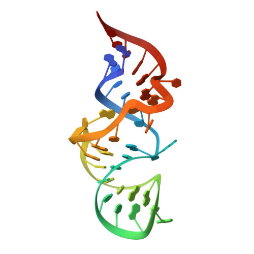
Recognition of planar and nonplanar ligands in the malachite green-RNA aptamer complex.
Flinders, J., DeFina, S.C., Brackett, D.M., Baugh, C., Wilson, C., Dieckmann, T.(2004) Chembiochem 5: 62-72
- PubMed: 14695514
- DOI: https://doi.org/10.1002/cbic.200300701
- Primary Citation of Related Structures:
1Q8N - PubMed Abstract:
Ribonucleic acids are an attractive drug target owing to their central role in many pathological processes. Notwithstanding this potential, RNA has only rarely been successfully targeted with novel drugs. The difficulty of targeting RNA is at least in part due to the unusual mode of binding found in most small-molecule-RNA complexes: the ligand binding pocket of the RNA is largely unstructured in the absence of ligand and forms a defined structure only with the ligand acting as scaffold for folding. Moreover, electrostatic interactions between RNA and ligand can also induce significant changes in the ligand structure due to the polyanionic nature of the RNA. Aptamers are ideal model systems to study these kinds of interactions owing to their small size and the ease with which they can be evolved to recognize a large variety of different ligands. Here we present the solution structure of an RNA aptamer that binds triphenyl dyes in complex with malachite green and compare it with a previously determined crystal structure of a complex formed with tetramethylrosamine. The structures illustrate how the same RNA binding pocket can adapt to accommodate both planar and nonplanar ligands. Binding studies with single- and double-substitution mutant aptamers are used to correlate three-dimensional structure with complex stability. The two RNA-ligand complex structures allow a discussion of structural changes that have been observed in the ligand in the context of the overall complex structure. Base pairing and stacking interactions within the RNA fold the phosphate backbone into a structure that results in an asymmetric charge distribution within the binding pocket that forces the ligand to adapt through a redistribution of the positive partial charge.
Organizational Affiliation:
Department of Chemistry, University of California, Davis, CA 95616, USA.


