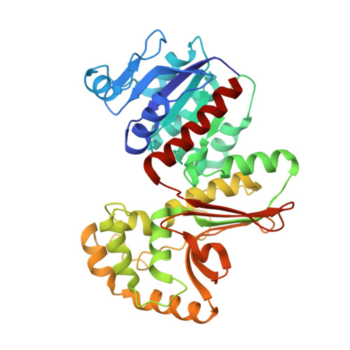
1Q7G

Homoserine Dehydrogenase in complex with suicide inhibitor complex NAD-5-hydroxy-4-Oxonorvaline


Experimental Data Snapshot

  • Method: X-RAY DIFFRACTION
  • Resolution: 2.60 Å
  • R-Value Free: 0.306 
  • R-Value Work: 0.247 
  • R-Value Observed: 0.247 

Starting Model: experimental
View more details

wwPDB Validation   3D Report Full Report


Ligand Structure Quality Assessment 


This is version 1.3 of the entry. See complete history


Literature

Enzyme assisted suicide: Molecular basis for the antifungal activity of 5-hydroxy-4-oxonorvaline by potent inhibition of homoserine dehydrogenase

Jacques, S.L.Mirza, I.A.Ejim, L.Koteva, K.Hughes, D.W.Green, K.Kinach, R.Honek, J.F.Lai, H.K.Berghuis, A.M.Wright, G.D.

(2003) Chem Biol 10: 989-995

  • DOI: https://doi.org/10.1016/j.chembiol.2003.09.015
  • Primary Citation of Related Structures:  
    1Q7G

  • PubMed Abstract: 

    The structure of the antifungal drug 5-hydroxy-4-oxonorvaline (HON) in complex with its target homoserine dehydrogenase (HSD) has been determined by X-ray diffraction to 2.6 A resolution. HON shows potent in vitro and in vivo activity against various fungal pathogens despite its weak (2 mM) affinity for HSD in the steady state. The structure together with structure-activity relationship studies, mass spectrometry experiments, and spectroscopic data reveals that the molecular mechanism of antifungal action conferred by HON involves enzyme-dependent formation of a covalent adduct between C4 of the nicotinamide ring of NAD(+) and C5 of HON. Furthermore, novel interactions are involved in stabilizing the (HON*NAD)-adduct, which are not observed in the enzyme's ternary complex structure. These findings clarify the apparent paradox of the potent antifungal actions of HON given its weak steady-state inhibition characteristics.


  • Organizational Affiliation

    Antimicrobial Research Centre and Department of Biochemistry, McMaster University, Hamilton L8N 3Z5, Canada.


Macromolecules
Find similar proteins by:  (by identity cutoff)  |  3D Structure
Entity ID: 1
MoleculeChains Sequence LengthOrganismDetailsImage
Homoserine dehydrogenase
A, B
359Saccharomyces cerevisiaeMutation(s): 0 
Gene Names: HOM6YJR139C OR J2132
EC: 1.1.1.3
UniProt
Find proteins for P31116 (Saccharomyces cerevisiae (strain ATCC 204508 / S288c))
Explore P31116 
Go to UniProtKB:  P31116
Entity Groups  
Sequence Clusters30% Identity50% Identity70% Identity90% Identity95% Identity100% Identity
UniProt GroupP31116
Sequence Annotations
Expand
  • Reference Sequence
Experimental Data & Validation

Experimental Data

  • Method: X-RAY DIFFRACTION
  • Resolution: 2.60 Å
  • R-Value Free: 0.306 
  • R-Value Work: 0.247 
  • R-Value Observed: 0.247 
  • Space Group: P 43 21 2
Unit Cell:
Length ( Å )Angle ( ˚ )
a = 80.863α = 90
b = 80.863β = 90
c = 248.986γ = 90
Software Package:
Software NamePurpose
CNSrefinement
SCALEPACKdata scaling
CNSphasing

Structure Validation

View Full Validation Report


Ligand Structure Quality Assessment 


Entry History 

Deposition Data

Revision History  (Full details and data files)

  • Version 1.0: 2003-10-21
    Type: Initial release
  • Version 1.1: 2008-04-29
    Changes: Version format compliance
  • Version 1.2: 2011-07-13
    Changes: Version format compliance
  • Version 1.3: 2023-08-16
    Changes: Data collection, Database references, Derived calculations, Refinement description