1Q57 | pdb_00001q57

The Crystal Structure of the Bifunctional Primase-Helicase of Bacteriophage T7


Experimental Data Snapshot

  • Method: X-RAY DIFFRACTION
  • Resolution: 3.45 Å
  • R-Value Free: 
    0.326 (Depositor), 0.311 (DCC) 
  • R-Value Work: 
    0.299 (Depositor), 0.284 (DCC) 
  • R-Value Observed: 
    0.301 (Depositor) 

wwPDB Validation 3D Report Full Report

Validation slider image for 1Q57

This is version 1.4 of the entry. See complete history

Literature

The Crystal Structure of the Bifunctional Primase-Helicase of Bacteriophage T7

Toth, E.A.Li, Y.Sawaya, M.R.Cheng, Y.Ellenberger, T.

(2003) Mol Cell 12: 1113-1123

  • DOI: https://doi.org/10.1016/s1097-2765(03)00442-8
  • Primary Citation Related Structures: 
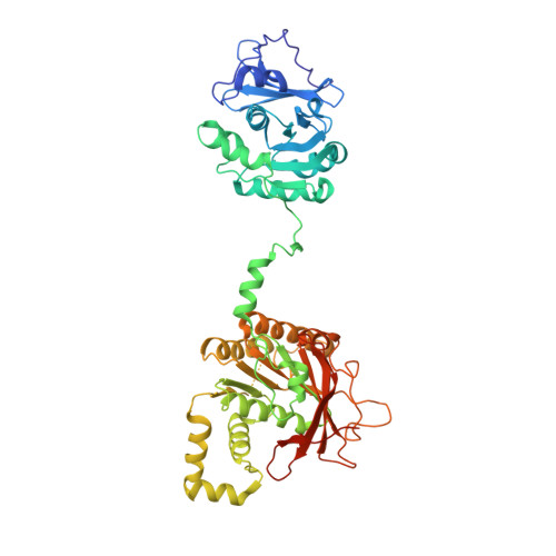
    1Q57

  • PubMed Abstract: 

    Within minutes after infecting Escherichia coli, bacteriophage T7 synthesizes many copies of its genomic DNA. The lynchpin of the T7 replication system is a bifunctional primase-helicase that unwinds duplex DNA at the replication fork while initiating the synthesis of Okazaki fragments on the lagging strand. We have determined a 3.45 A crystal structure of the T7 primase-helicase that shows an articulated arrangement of the primase and helicase sites. The crystallized primase-helicase is a heptamer with a crown-like shape, reflecting an intimate packing of helicase domains into a ring that is topped with loosely arrayed primase domains. This heptameric isoform can accommodate double-stranded DNA in its central channel, which nicely explains its recently described DNA remodeling activity. The double-jointed structure of the primase-helicase permits a free range of motion for the primase and helicase domains that suggests how the continuous unwinding of DNA at the replication fork can be periodically coupled to Okazaki fragment synthesis.


  • Organizational Affiliation
    • Department of Biological Chemistry and Molecular Pharmacology, Harvard Medical School, 240 Longwood Avenue, Boston, MA 02115, USA.

Macromolecule Content 

  • Total Structure Weight: 391 kDa 
  • Atom Count: 26,208 
  • Modeled Residue Count: 3,381 
  • Deposited Residue Count: 3,521 
  • Unique protein chains: 1

Macromolecules

Find similar proteins by:|  3D Structure
Entity ID: 1
MoleculeChains  Sequence LengthOrganismDetailsImage
DNA primase/helicase
A, B, C, D, E
A, B, C, D, E, F, G
503Escherichia phage T7Mutation(s): 2 
Gene Names: 4
EC: 2.7.7 (PDB Primary Data), 5.6.2.3 (UniProt)
UniProt
Find proteins for P03692 (Escherichia phage T7)
Explore P03692 
Go to UniProtKB:  P03692
Entity Groups
Sequence Clusters30% Identity50% Identity70% Identity90% Identity95% Identity100% Identity
UniProt GroupP03692
Sequence Annotations
Expand
Reference Sequence

Experimental Data & Validation

Experimental Data

  • Method: X-RAY DIFFRACTION
  • Resolution: 3.45 Å
  • R-Value Free:  0.326 (Depositor), 0.311 (DCC) 
  • R-Value Work:  0.299 (Depositor), 0.284 (DCC) 
  • R-Value Observed: 0.301 (Depositor) 
Space Group: P 1 21 1
Unit Cell:
Length ( Å )Angle ( ˚ )
a = 117.179α = 90
b = 171.567β = 99.86
c = 118.581γ = 90
Software Package:
Software NamePurpose
REFMACrefinement
HKL-2000data reduction
SCALEPACKdata scaling
SHARPphasing

Structure Validation

View Full Validation Report


Entry History 

Deposition Data

Revision History  (Full details and data files)

  • Version 1.0: 2003-11-25
    Type: Initial release
  • Version 1.1: 2008-04-29
    Changes: Version format compliance
  • Version 1.2: 2011-07-13
    Changes: Advisory, Version format compliance
  • Version 1.3: 2021-10-27
    Changes: Database references
  • Version 1.4: 2024-02-14
    Changes: Data collection, Refinement description