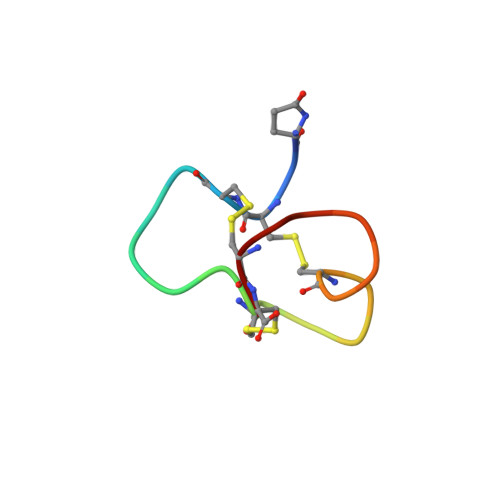
Structural basis for tetrodotoxin-resistant sodium channel binding by mu-conotoxin SmIIIA.
Keizer, D.W., West, P.J., Lee, E.F., Yoshikami, D., Olivera, B.M., Bulaj, G., Norton, R.S.(2003) J Biological Chem 278: 46805-46813
- PubMed: 12970353 Search on PubMed
- DOI: https://doi.org/10.1074/jbc.M309222200
- Primary Citation Related Structures:
1Q2J - PubMed Abstract:
SmIIIA is a new micro-conotoxin isolated recently from Conus stercusmuscarum. Although it shares several biochemical characteristics with other micro-conotoxins (the arrangement of cysteine residues and a conserved arginine believed to interact with residues near the channel pore), it has several distinctive features, including the absence of hydroxyproline, and is the first specific antagonist of tetrodotoxin-resistant voltage-gated sodium channels to be characterized. It therefore represents a potentially useful tool to investigate the functional roles of these channels. We have determined the three-dimensional structure of SmIIIA in aqueous solution. Consistent with the absence of hydroxyprolines, SmIIIA adopts a single conformation with all peptide bonds in the trans configuration. The spatial orientations of several conserved Arg and Lys side chains, including Arg14 (using a consensus numbering system), which plays a key role in sodium channel binding, are similar to those in other micro-conotoxins but the N-terminal regions differ, reflecting the trans conformation for the peptide bond preceding residue 8 in SmIIIA, as opposed to the cis conformation in micro-conotoxins GIIIA and GIIIB. Comparison of the surfaces of SmIIIA with other micro-conotoxins suggests that the affinity of SmIIIA for TTX-resistant channels is influenced by the Trp15 side chain, which is unique to SmIIIA. Arg17, which replaces Lys in the other micro-conotoxins, may also be important. Consistent with these inferences from the structure, assays of two chimeras of SmIIIA and PIIIA in which their N- and C-terminal halves were recombined, indicated that residues in the C-terminal half of SmIIIA confer affinity for tetrodotoxin-resistant sodium channels in the cell bodies of frog sympathetic neurons. SmIIIA and the chimera possessing the C-terminal half of SmIIIA also inhibit tetrodotoxin-resistant sodium channels in the postganglionic axons of sympathetic neurons, as indicated by their inhibition of C-neuron compound action potentials that persist in the presence of tetrodotoxin.
- The Walter & Eliza Hall Institute of Medical Research, 1G Royal Parade, Parkville, Victoria 3050, Australia.
Organizational Affiliation: