1PSA | pdb_00001psa

STRUCTURE OF A PEPSIN(SLASH)RENIN INHIBITOR COMPLEX REVEALS A NOVEL CRYSTAL PACKING INDUCED BY MINOR CHEMICAL ALTERATIONS IN THE INHIBITOR


Experimental Data Snapshot

  • Method: X-RAY DIFFRACTION
  • Resolution: 2.90 Å
  • R-Value Work: 
    0.139 (Depositor), 0.129 (DCC) 

wwPDB Validation 3D Report Full Report

Validation slider image for 1PSA

Ligand Structure Quality Assessment 


This is version 1.5 of the entry. See complete history

Literature

Structure of a pepsin/renin inhibitor complex reveals a novel crystal packing induced by minor chemical alterations in the inhibitor.

Chen, L.Erickson, J.W.Rydel, T.J.Park, C.H.Neidhart, D.Luly, J.Abad-Zapatero, C.

(1992) Acta Crystallogr B 48: 476-488

  • DOI: https://doi.org/10.1107/s0108768192001939
  • Primary Citation Related Structures: 
    1PSA

  • PubMed Abstract: 

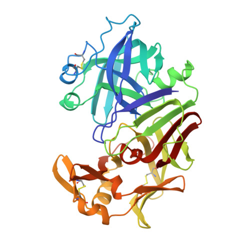
    The structure determination by molecular replacement methods of a monoclinic pepsin/renin inhibitor complex crystal, with two molecules in the asymmetric unit, is presented. The atomic model, consisting of two liganded pepsin molecules and 110 water molecules, has been refined to a final crystallographic R value of 0.139 for data between 8 and 2.9 A resolution. The structure reveals a previously undescribed pepsin dimer formed predominantly by polar interactions. Inhibitor binding induces global structural changes in the native enzyme similar, but not identical, to the ones observed in other chemically similar pepsin/renin inhibitor complexes crystallized in an orthorhombic form. A region of the polypeptide chain (residues 292-297) which was not visible in the orthorhombic crystal is well ordered in the presently described structure; possibly induced by crystal contacts. The crystal packing of native pepsin is compared with the two different crystal forms of the inhibited enzyme.


  • Organizational Affiliation
    • Laboratory of Protein Crystallography, Abbott Laboratories, Abbott Park, IL 60064.

Macromolecule Content 

  • Total Structure Weight: 70.11 kDa 
  • Atom Count: 5,044 
  • Modeled Residue Count: 652 
  • Deposited Residue Count: 652 
  • Unique protein chains: 1

Macromolecules

Find similar proteins by:|  3D Structure
Entity ID: 1
MoleculeChains  Sequence LengthOrganismDetailsImage
PEPSIN A
A, B
326Sus scrofaMutation(s): 0 
EC: 3.4.23.1
UniProt
Find proteins for P00791 (Sus scrofa)
Explore P00791 
Go to UniProtKB:  P00791
Entity Groups
Sequence Clusters30% Identity50% Identity70% Identity90% Identity95% Identity100% Identity
UniProt GroupP00791
Sequence Annotations
Expand
Reference Sequence

Small Molecules

Ligands 1 Unique
IDChains Name / Formula / InChI Key2D Diagram3D Interactions
0ZL

Query on 0ZL


Download:Ideal Coordinates CCD File
C [auth A],
D [auth B]
N-(ethoxycarbonyl)-L-leucyl-N-[(1R,2S,3S)-1-(cyclohexylmethyl)-2,3-dihydroxy-5-methylhexyl]-L-leucinamide
C29 H55 N3 O6
OAFXIYWBKGSXMT-WBAQKLHDSA-N

Experimental Data & Validation

Experimental Data

  • Method: X-RAY DIFFRACTION
  • Resolution: 2.90 Å
  • R-Value Work:  0.139 (Depositor), 0.129 (DCC) 
Space Group: P 1 1 21
Unit Cell:
Length ( Å )Angle ( ˚ )
a = 54.1α = 90
b = 74.4β = 90
c = 76.5γ = 100.8
Software Package:
Software NamePurpose
X-PLORrefinement

Structure Validation

View Full Validation Report


Ligand Structure Quality Assessment 


Entry History 

Deposition Data

Revision History  (Full details and data files)

  • Version 1.0: 1994-01-31
    Type: Initial release
  • Version 1.1: 2008-03-24
    Changes: Version format compliance
  • Version 1.2: 2011-07-13
    Changes: Atomic model, Database references, Derived calculations, Non-polymer description, Structure summary, Version format compliance
  • Version 1.3: 2012-12-12
    Changes: Other
  • Version 1.4: 2017-11-29
    Changes: Advisory, Derived calculations, Other
  • Version 1.5: 2024-10-30
    Changes: Advisory, Data collection, Database references, Derived calculations, Structure summary