1PEA | pdb_00001pea

AMIDE RECEPTOR/NEGATIVE REGULATOR OF THE AMIDASE OPERON OF PSEUDOMONAS AERUGINOSA (AMIC) COMPLEXED WITH ACETAMIDE


Experimental Data Snapshot

  • Method: X-RAY DIFFRACTION
  • Resolution: 2.10 Å
  • R-Value Free: 
    0.275 (Depositor) 
  • R-Value Work: 
    0.206 (Depositor) 
  • R-Value Observed: 
    0.206 (Depositor) 

wwPDB Validation 3D Report Full Report

Validation slider image for 1PEA

This is version 1.3 of the entry. See complete history

Literature

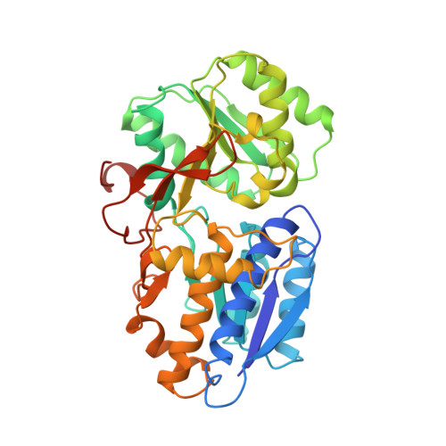
Crystal structure of AmiC: the controller of transcription antitermination in the amidase operon of Pseudomonas aeruginosa.

Pearl, L.O'Hara, B.Drew, R.Wilson, S.

(1994) EMBO J 13: 5810-5817

  • DOI: https://doi.org/10.1002/j.1460-2075.1994.tb06924.x
  • Primary Citation Related Structures: 
    1PEA

  • PubMed Abstract: 

    The crystal structure for the negative regulator (AmiC) of the amidase operon from Pseudomonas aeruginosa has been solved at a resolution of 2.1 A. AmiC is the amide sensor protein in the amidase operon and regulates the activity of the transcription antitermination factor AmiR, which in turn regulates amidase expression. The AmiC structure consists of two domains with an alternating beta-alpha-beta topology. The two domains are separated by a central cleft and the amide binding site is positioned in this cleft at the interface of the domains. The overall fold for AmiC is extremely similar to that for the leucine-isoleucine-valine binding protein (LivJ) of Escherichia coli despite only 17% sequence identity, however, the two domains of AmiC are substantially closed compared with LivJ. The closed structure of AmiC is stabilized significantly by the bound acetamide, suggesting a molecular mechanism for the process of amide induction. The amide binding site is extremely specific for acetamide and would not allow a closed conformation in the presence of the anti-inducer molecule butyramide.


  • Organizational Affiliation
    • Department of Biochemistry and Molecular Biology, University College London, UK.

Macromolecule Content 

  • Total Structure Weight: 43.02 kDa 
  • Atom Count: 3,044 
  • Modeled Residue Count: 368 
  • Deposited Residue Count: 385 
  • Unique protein chains: 1

Macromolecules

Find similar proteins by:|  3D Structure
Entity ID: 1
MoleculeChains  Sequence LengthOrganismDetailsImage
AMIDASE OPERON385Pseudomonas aeruginosaMutation(s): 0 
UniProt
Find proteins for P27017 (Pseudomonas aeruginosa (strain ATCC 15692 / DSM 22644 / CIP 104116 / JCM 14847 / LMG 12228 / 1C / PRS 101 / PAO1))
Explore P27017 
Go to UniProtKB:  P27017
Entity Groups
Sequence Clusters30% Identity50% Identity70% Identity90% Identity95% Identity100% Identity
UniProt GroupP27017
Sequence Annotations
Expand
Reference Sequence

Small Molecules

Ligands 1 Unique
IDChains Name / Formula / InChI Key2D Diagram3D Interactions
ACM

Query on ACM


Download:Ideal Coordinates CCD File
B [auth A]ACETAMIDE
C2 H5 N O
DLFVBJFMPXGRIB-UHFFFAOYSA-N

Experimental Data & Validation

Experimental Data

  • Method: X-RAY DIFFRACTION
  • Resolution: 2.10 Å
  • R-Value Free:  0.275 (Depositor) 
  • R-Value Work:  0.206 (Depositor) 
  • R-Value Observed: 0.206 (Depositor) 
Space Group: P 42 21 2
Unit Cell:
Length ( Å )Angle ( ˚ )
a = 104.96α = 90
b = 104.96β = 90
c = 65.8γ = 90
Software Package:
Software NamePurpose
X-PLORmodel building
X-PLORrefinement
MOSFLMdata reduction
X-PLORphasing

Structure Validation

View Full Validation Report


Entry History 

Deposition Data

Revision History  (Full details and data files)

  • Version 1.0: 1996-04-03
    Type: Initial release
  • Version 1.1: 2008-03-03
    Changes: Version format compliance
  • Version 1.2: 2011-07-13
    Changes: Version format compliance
  • Version 1.3: 2024-02-14
    Changes: Data collection, Database references, Derived calculations, Other