1PE3 | pdb_00001pe3

Solution structure of the disulphide-linked dimer of human intestinal trefoil factor (TFF3)


Experimental Data Snapshot

  • Method: SOLUTION NMR
  • Conformers Calculated: 100 
  • Conformers Submitted: 47 
  • Selection Criteria: structures with acceptable covalent geometry, structures with favorable non-bond energy, structures with the least restraint violations, structures with the lowest energy, target function 

wwPDB Validation 3D Report Full Report

Validation slider image for 1PE3

This is version 1.4 of the entry. See complete history

Literature

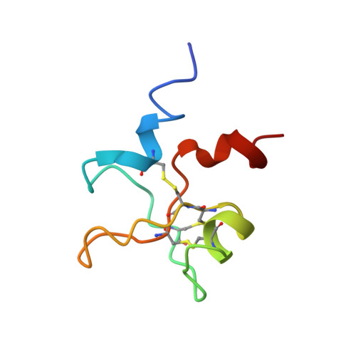
Solution structure of the disulfide-linked dimer of human intestinal trefoil factor (TFF3): the intermolecular orientation and interactions are markedly different from those of other dimeric trefoil proteins.

Muskett, F.W.May, F.E.Westley, B.R.Feeney, J.

(2003) Biochemistry 42: 15139-15147

  • DOI: https://doi.org/10.1021/bi030182k
  • Primary Citation Related Structures: 
    1PE3

  • PubMed Abstract: 

    The trefoil protein TFF3 forms a homodimer (via a disulfide linkage) that is thought to have increased biological activity over the monomer. The solution structure of the TFF3 dimer has been determined by NMR and compared with the structure of the TFF3 monomer and with other trefoil dimer structures (TFF1 and TFF2). The most significant structural differences between the trefoil domain in the monomer and dimer TFF3 are in the orientations of the N-terminal 3(10)-helix (residues 10-12) and in the presence in the dimer of an additional 3(10)-helix (residues 53-55) outside of the core region. The TFF3 dimer forms a more compact structure as compared with the TFF1 dimer where the two trefoil domains are connected by a flexible region with the monomer units being at variable distances from each other and in many different orientations. Although TFF2 is also a compact structure, the dispositions of its monomer units are very different from those of TFF3. The structural differences between the dimers result in the two putative receptor/ligand binding sites that remain solvent exposed in the dimeric structures having very different dispositions in the different dimers. Such differences have significant implications for the mechanism of action and functional specificity for the TFF class of proteins.


  • Organizational Affiliation
    • MRC Biomedical NMR Centre, National Institute for Medical Research, Mill Hill, London NW7 1AA, United Kingdom. fwm1@leicester.ac.uk

Macromolecule Content 

  • Total Structure Weight: 13.17 kDa 
  • Atom Count: 914 
  • Modeled Residue Count: 118 
  • Deposited Residue Count: 118 
  • Unique protein chains: 1

Macromolecules

Find similar proteins by:|  3D Structure
Entity ID: 1
MoleculeChains  Sequence LengthOrganismDetailsImage
Trefoil factor 3A [auth 1],
B [auth 2]
59Homo sapiensMutation(s): 0 
Gene Names: TFF3 OR ITF OR TFI
UniProt & NIH Common Fund Data Resources
Find proteins for Q07654 (Homo sapiens)
Explore Q07654 
Go to UniProtKB:  Q07654
PHAROS:  Q07654
GTEx:  ENSG00000160180 
Entity Groups
Sequence Clusters30% Identity50% Identity70% Identity90% Identity95% Identity100% Identity
UniProt GroupQ07654
Sequence Annotations
Expand
Reference Sequence

Experimental Data & Validation

Experimental Data

  • Method: SOLUTION NMR
  • Conformers Calculated: 100 
  • Conformers Submitted: 47 
  • Selection Criteria: structures with acceptable covalent geometry, structures with favorable non-bond energy, structures with the least restraint violations, structures with the lowest energy, target function 

Structure Validation

View Full Validation Report


Entry History 

Deposition Data

Revision History  (Full details and data files)

  • Version 1.0: 2004-03-09
    Type: Initial release
  • Version 1.1: 2008-04-29
    Changes: Version format compliance
  • Version 1.2: 2011-07-13
    Changes: Version format compliance
  • Version 1.3: 2022-02-23
    Changes: Database references, Derived calculations
  • Version 1.4: 2024-11-20
    Changes: Data collection, Structure summary