1P9B | pdb_00001p9b

Structure of fully ligated Adenylosuccinate synthetase from Plasmodium falciparum


Experimental Data Snapshot

  • Method: X-RAY DIFFRACTION
  • Resolution: 2.00 Å
  • R-Value Free: 
    0.241 (Depositor), 0.244 (DCC) 
  • R-Value Work: 
    0.199 (Depositor), 0.200 (DCC) 

Starting Model: experimental
View more details

wwPDB Validation 3D Report Full Report

Validation slider image for 1P9B

Ligand Structure Quality Assessment 


This is version 1.3 of the entry. See complete history

Literature

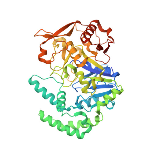
Crystal Structure of Fully Ligated Adenylosuccinate Synthetase from Plasmodium falciparum.

Eaazhisai, K.Jayalakshmi, R.Gayathri, P.Anand, R.P.Sumathy, K.Balaram, H.Murthy, M.R.

(2004) J Mol Biology 335: 1251-1264

  • DOI: https://doi.org/10.1016/j.jmb.2003.11.036
  • Primary Citation Related Structures: 
    1P9B

  • PubMed Abstract: 

    In the absence of the de novo purine nucleotide biosynthetic pathway in parasitic protozoa, purine salvage is of primary importance for parasite survival. Enzymes of the salvage pathway are, therefore, good targets for anti-parasitic drugs. Adenylosuccinate synthetase (AdSS), catalysing the first committed step in the synthesis of AMP from IMP, is a potential target for anti-protozoal chemotherapy. We report here the crystal structure of adenylosuccinate synthetase from the malaria parasite, Plasmodium falciparum, complexed to 6-phosphoryl IMP, GDP, Mg2+ and the aspartate analogue, hadacidin at 2 A resolution. The overall architecture of P. falciparum AdSS (PfAdSS) is similar to the known structures from Escherichia coli, mouse and plants. Differences in substrate interactions seen in this structure provide a plausible explanation for the kinetic differences between PfAdSS and the enzyme from other species. Additional hydrogen bonding interactions of the protein with GDP may account for the ordered binding of substrates to the enzyme. The dimer interface of PfAdSS is also different, with a pronounced excess of positively charged residues. Differences highlighted here provide a basis for the design of species-specific inhibitors of the enzyme.


  • Organizational Affiliation
    • Molecular Biophysics Unit, UGC Centre of Advanced Study, Indian Institute of Science, Bangalore 560012, India.

Macromolecule Content 

  • Total Structure Weight: 51.17 kDa 
  • Atom Count: 3,697 
  • Modeled Residue Count: 424 
  • Deposited Residue Count: 442 
  • Unique protein chains: 1

Macromolecules

Find similar proteins by:|  3D Structure
Entity ID: 1
MoleculeChains  Sequence LengthOrganismDetailsImage
Adenylosuccinate Synthetase442Plasmodium falciparumMutation(s): 0 
EC: 6.3.4.4
UniProt
Find proteins for Q9U8D3 (Plasmodium falciparum)
Explore Q9U8D3 
Go to UniProtKB:  Q9U8D3
Entity Groups
Sequence Clusters30% Identity50% Identity70% Identity90% Identity95% Identity100% Identity
UniProt GroupQ9U8D3
Sequence Annotations
Expand
Reference Sequence

Small Molecules

Ligands 5 Unique
IDChains Name / Formula / InChI Key2D Diagram3D Interactions
GDP

Query on GDP


Download:Ideal Coordinates CCD File
F [auth A]GUANOSINE-5'-DIPHOSPHATE
C10 H15 N5 O11 P2
QGWNDRXFNXRZMB-UUOKFMHZSA-N
IMO

Query on IMO


Download:Ideal Coordinates CCD File
D [auth A]6-O-PHOSPHORYL INOSINE MONOPHOSPHATE
C10 H14 N4 O11 P2
RXRZOKQPANIEDW-KQYNXXCUSA-N
HDA

Query on HDA


Download:Ideal Coordinates CCD File
E [auth A]HADACIDIN
C3 H5 N O4
URJHVPKUWOUENU-UHFFFAOYSA-N
NO3

Query on NO3


Download:Ideal Coordinates CCD File
C [auth A]NITRATE ION
N O3
NHNBFGGVMKEFGY-UHFFFAOYSA-N
MG

Query on MG


Download:Ideal Coordinates CCD File
B [auth A]MAGNESIUM ION
Mg
JLVVSXFLKOJNIY-UHFFFAOYSA-N

Experimental Data & Validation

Experimental Data

  • Method: X-RAY DIFFRACTION
  • Resolution: 2.00 Å
  • R-Value Free:  0.241 (Depositor), 0.244 (DCC) 
  • R-Value Work:  0.199 (Depositor), 0.200 (DCC) 
Space Group: C 2 2 21
Unit Cell:
Length ( Å )Angle ( ˚ )
a = 91.579α = 90
b = 117.117β = 90
c = 80.416γ = 90
Software Package:
Software NamePurpose
DENZOdata reduction
SCALEPACKdata scaling
AMoREphasing
CNSrefinement

Structure Validation

View Full Validation Report


Ligand Structure Quality Assessment 


Entry History 

Deposition Data

Revision History  (Full details and data files)

  • Version 1.0: 2004-02-03
    Type: Initial release
  • Version 1.1: 2008-04-29
    Changes: Version format compliance
  • Version 1.2: 2011-07-13
    Changes: Derived calculations, Version format compliance
  • Version 1.3: 2023-10-25
    Changes: Data collection, Database references, Derived calculations, Refinement description