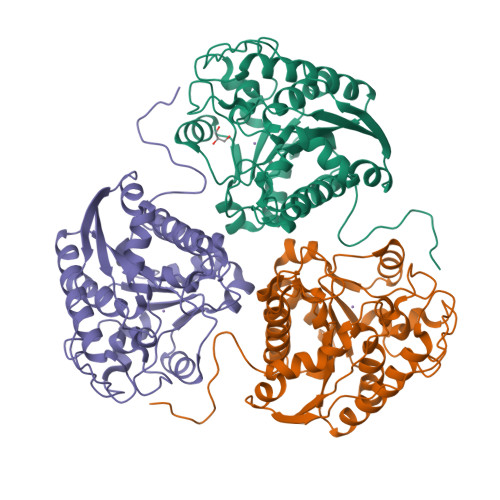
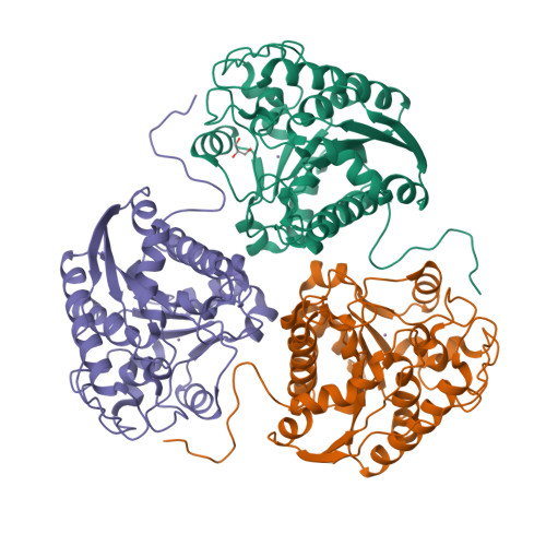
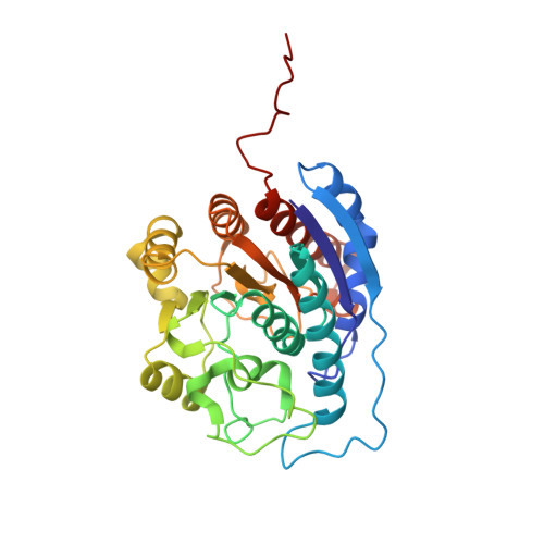
1P8N

Structural and Functional Importance of First-Shell Metal Ligands in the Binuclear Manganese Cluster of Arginase I.


Experimental Data Snapshot

  • Method: X-RAY DIFFRACTION
  • Resolution: 2.90 Å
  • R-Value Free: 0.305 
  • R-Value Work: 0.286 
  • R-Value Observed: 0.286 

Starting Model: experimental
View more details

wwPDB Validation   3D Report Full Report


This is version 1.4 of the entry. See complete history


Literature

Structural and functional importance of first-shell metal ligands in the binuclear manganese cluster of arginase I

Cama, E.Emig, F.A.Ash, D.E.Christianson, D.W.

(2003) Biochemistry 42: 7748-7758

  • DOI: https://doi.org/10.1021/bi030074y
  • Primary Citation of Related Structures:  
    1P8M, 1P8N, 1P8O, 1P8P, 1P8Q, 1P8R, 1P8S

  • PubMed Abstract: 

    Arginase is a binuclear manganese metalloenzyme that hydrolyzes l-arginine to form l-ornithine and urea. The three-dimensional structures of D128E, D128N, D232A, D232C, D234E, H101N, and H101E arginases I have been determined by X-ray crystallographic methods to elucidate the roles of the first-shell metal ligands in the stability and catalytic activity of the enzyme. This work represents the first structure-based dissection of the binuclear manganese cluster using site-directed mutagenesis and X-ray crystallography. Substitution of the metal ligands compromises the catalytic activity of the enzyme, either by the loss or disruption of the metal cluster or the nucleophilic metal-bridging hydroxide ion. However, the substitution of the metal ligands or the reduction of Mn(2+)(A) or Mn(2+)(B) occupancy does not compromise enzyme-substrate affinity as reflected by K(M), which remains relatively invariant across this series of arginase variants. This implicates a nonmetal binding site for substrate l-arginine in the precatalytic Michaelis complex, as proposed based on analysis of the native enzyme structure (Kanyo, Z. F., Scolnick, L. R., Ash, D. E., and Christianson, D. W. (1996) Nature 383, 554-557).


  • Organizational Affiliation

    Roy and Diana Vagelos Laboratories, Department of Chemistry, University of Pennsylvania, Philadelphia, Pennsylvania 19104-6323, USA.


Macromolecules
Find similar proteins by:  (by identity cutoff)  |  3D Structure
Entity ID: 1
MoleculeChains Sequence LengthOrganismDetailsImage
Arginase 1
A, B, C
314Rattus norvegicusMutation(s): 1 
Gene Names: ARG1
EC: 3.5.3.1
UniProt
Find proteins for P07824 (Rattus norvegicus)
Explore P07824 
Go to UniProtKB:  P07824
Entity Groups  
Sequence Clusters30% Identity50% Identity70% Identity90% Identity95% Identity100% Identity
UniProt GroupP07824
Sequence Annotations
Expand
  • Reference Sequence
Experimental Data & Validation

Experimental Data

  • Method: X-RAY DIFFRACTION
  • Resolution: 2.90 Å
  • R-Value Free: 0.305 
  • R-Value Work: 0.286 
  • R-Value Observed: 0.286 
  • Space Group: P 32
Unit Cell:
Length ( Å )Angle ( ˚ )
a = 90.337α = 90
b = 90.337β = 90
c = 104.985γ = 120
Software Package:
Software NamePurpose
CNSrefinement
DENZOdata reduction
SCALEPACKdata scaling
AMoREphasing

Structure Validation

View Full Validation Report


Entry History 

Deposition Data

Revision History  (Full details and data files)

  • Version 1.0: 2003-06-17
    Type: Initial release
  • Version 1.1: 2008-04-29
    Changes: Version format compliance
  • Version 1.2: 2011-07-13
    Changes: Non-polymer description, Version format compliance
  • Version 1.3: 2021-10-27
    Changes: Database references, Derived calculations
  • Version 1.4: 2023-08-16
    Changes: Data collection, Refinement description