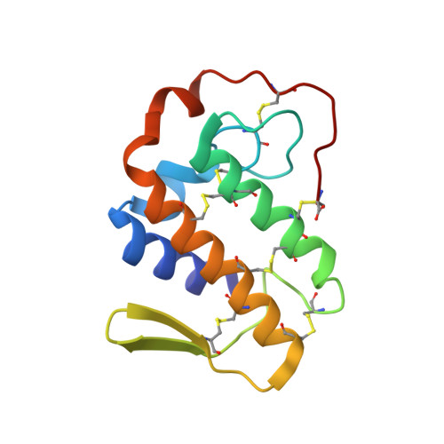
Structure of an acidic phospholipase A2 from Indian saw-scaled viper (Echis carinatus) at 2.6 A resolution reveals a novel intermolecular interaction.
Jasti, J., Paramasivam, M., Srinivasan, A., Singh, T.P.(2004) Acta Crystallogr D Biol Crystallogr 60: 66-72
- PubMed: 14684894
- DOI: https://doi.org/10.1107/s090744490302208x
- Primary Citation of Related Structures:
1OZ6 - PubMed Abstract:
The crystal structure of an acidic phospholipase A(2) from the venom of Echis carinatus (saw-scaled viper; scPLA(2)) has been determined at 2.6 A resolution and refined to a crystallographic R factor of 0.192. Although the overall structure of scPLA(2) is essentially similar to those of other group II acidic PLA(2)s from different species, it shows unique features in several parts. Particularly noteworthy is the C-terminal part, which folds differently to those of other group II PLA(2)s. This part is considered to be responsible for inhibition of the platelet-aggregation activity. The calcium-binding loop is tightly organized with sevenfold coordination. Another striking feature of scPLA(2) is the involvement of Asn79 O(delta1) of a symmetry-related molecule in a coordination linkage with Ca(2+) of the calcium-binding loop. This is the first observation of an internal metal ion participating in an intermolecular interaction. The beta-wing of a molecule is deeply inserted into the hydrophobic channel of another molecule and forms several intermolecular interactions. This results in the formation of an infinite chain of molecules. These chains are stacked in an antiparallel arrangement in the crystals.
- Department of Biophysics, All India Institute of Medical Sciences, New Delhi 110 029, India.
Organizational Affiliation:


