1OYP

Crystal Structure of the phosphorolytic exoribonuclease RNase PH from Bacillus subtilis


Experimental Data Snapshot

  • Method: X-RAY DIFFRACTION
  • Resolution: 2.76 Å
  • R-Value Free: 0.305 
  • R-Value Work: 0.285 

wwPDB Validation   3D Report Full Report


This is version 1.3 of the entry. See complete history


Literature

Crystal structure of the phosphorolytic exoribonuclease RNase PH from Bacillus subtilis and implications for its quaternary structure and tRNA binding.

Harlow, L.S.Kadziola, A.Jensen, K.F.Larsen, S.

(2004) Protein Sci 13: 668-677

  • DOI: https://doi.org/10.1110/ps.03477004
  • Primary Citation of Related Structures:  
    1OYP, 1OYR, 1OYS

  • PubMed Abstract: 

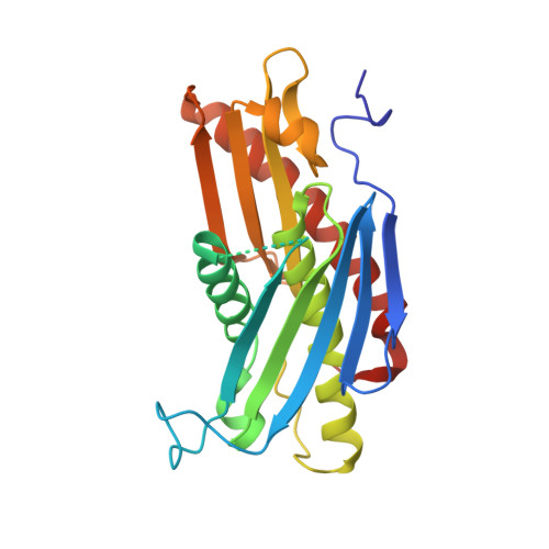
    RNase PH is a member of the family of phosphorolytic 3' --> 5' exoribonucleases that also includes polynucleotide phosphorylase (PNPase). RNase PH is involved in the maturation of tRNA precursors and especially important for removal of nucleotide residues near the CCA acceptor end of the mature tRNAs. Wild-type and triple mutant R68Q-R73Q-R76Q RNase PH from Bacillus subtilis have been crystallized and the structures determined by X-ray diffraction to medium resolution. Wild-type and triple mutant RNase PH crystallize as a hexamer and dimer, respectively. The structures contain a rare left-handed beta alpha beta-motif in the N-terminal portion of the protein. This motif has also been identified in other enzymes involved in RNA metabolism. The RNase PH structure and active site can, despite low sequence similarity, be overlayed with the N-terminal core of the structure and active site of Streptomyces antibioticus PNPase. The surface of the RNase PH dimer fit the shape of a tRNA molecule.


  • Organizational Affiliation

    Department of Biological Chemistry, University of Copenhagen, Universitetsparken 5, DK-2100 Copenhagen, Denmark.


Macromolecules
Find similar proteins by:  (by identity cutoff)  |  3D Structure
Entity ID: 1
MoleculeChains Sequence LengthOrganismDetailsImage
Ribonuclease PH
A, B, C, D, E
A, B, C, D, E, F
245Bacillus subtilisMutation(s): 0 
EC: 2.7.7.56
UniProt
Find proteins for P28619 (Bacillus subtilis (strain 168))
Explore P28619 
Go to UniProtKB:  P28619
Entity Groups  
Sequence Clusters30% Identity50% Identity70% Identity90% Identity95% Identity100% Identity
UniProt GroupP28619
Sequence Annotations
Expand
  • Reference Sequence
Small Molecules
Ligands 1 Unique
IDChains Name / Formula / InChI Key2D Diagram3D Interactions
SO4
Query on SO4

Download Ideal Coordinates CCD File 
G [auth A]
H [auth A]
I [auth B]
J [auth B]
K [auth C]
G [auth A],
H [auth A],
I [auth B],
J [auth B],
K [auth C],
L [auth C],
M [auth D],
N [auth D],
O [auth E],
P [auth E],
Q [auth F],
R [auth F]
SULFATE ION
O4 S
QAOWNCQODCNURD-UHFFFAOYSA-L
Experimental Data & Validation

Experimental Data

  • Method: X-RAY DIFFRACTION
  • Resolution: 2.76 Å
  • R-Value Free: 0.305 
  • R-Value Work: 0.285 
  • Space Group: P 21 21 21
Unit Cell:
Length ( Å )Angle ( ˚ )
a = 51.31α = 90
b = 163.56β = 90
c = 166.8γ = 90
Software Package:
Software NamePurpose
DENZOdata reduction
SCALEPACKdata scaling
AMoREphasing
CNSrefinement

Structure Validation

View Full Validation Report


Entry History 

Deposition Data

Revision History  (Full details and data files)

  • Version 1.0: 2004-03-09
    Type: Initial release
  • Version 1.1: 2008-04-29
    Changes: Version format compliance
  • Version 1.2: 2011-07-13
    Changes: Version format compliance
  • Version 1.3: 2024-02-14
    Changes: Data collection, Database references, Derived calculations