1OXE | pdb_00001oxe

Expansion of the Genetic Code Enables Design of a Novel "Gold" Class of Green Fluorescent Proteins


Experimental Data Snapshot

  • Method: X-RAY DIFFRACTION
  • Resolution: 1.15 Å
  • R-Value Free: 
    0.236 (Depositor) 
  • R-Value Work: 
    0.223 (Depositor) 
  • R-Value Observed: 
    0.223 (Depositor) 

wwPDB Validation 3D Report Full Report

Validation slider image for 1OXE

This is version 2.0 of the entry. See complete history

Literature

Expansion of the Genetic Code Enables Design of a Novel "Gold" Class of Green Fluorescent Proteins

Hyun Bae, J.Rubini, M.Jung, G.Wiegand, G.Seifert, M.H.Azim, M.K.Kim, J.S.Zumbusch, A.Holak, T.A.Moroder, L.Huber, R.Budisa, N.

(2003) J Mol Biology 328: 1071-1081

  • DOI: https://doi.org/10.1016/s0022-2836(03)00364-4
  • Primary Citation Related Structures: 
    1OXD, 1OXE, 1OXF

  • PubMed Abstract: 

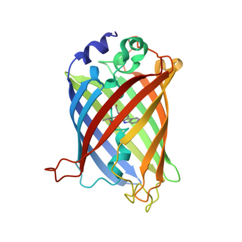
    Much effort has been dedicated to the design of significantly red shifted variants of the green fluorescent protein (GFP) from Aequoria victora (av). These approaches have been based on classical engineering with the 20 canonical amino acids. We report here an expansion of these efforts by incorporation of an amino substituted variant of tryptophan into the "cyan" GFP mutant, which turned it into a "gold" variant. This variant possesses a red shift in emission unprecedented for any avFP, similar to "red" FPs, but with enhanced stability and a very low aggregation tendency. An increasing number of non-natural amino acids are available for chromophore redesign (by engineering of the genetic code) and enable new general strategies to generate novel classes of tailor-made GFP proteins.


  • Organizational Affiliation
    • Max-Planck-Institut für Biochemie, Am Klopferspitz 18A, D-82152 Martinsried, Germany.

Macromolecule Content 

  • Total Structure Weight: 25.8 kDa 
  • Atom Count: 1,966 
  • Modeled Residue Count: 227 
  • Deposited Residue Count: 227 
  • Unique protein chains: 1

Macromolecules

Find similar proteins by:|  3D Structure
Entity ID: 1
MoleculeChains  Sequence LengthOrganismDetailsImage
cyan fluorescent protein cfp227cfp marker plasmid pWM1009Mutation(s): 2 

Small Molecules

Modified Residues  1 Unique
IDChains TypeFormula2D DiagramParent
CRF
Query on CRF
A
L-PEPTIDE LINKINGC17 H18 N4 O4THR, TRP, GLY

Experimental Data & Validation

Experimental Data

  • Method: X-RAY DIFFRACTION
  • Resolution: 1.15 Å
  • R-Value Free:  0.236 (Depositor) 
  • R-Value Work:  0.223 (Depositor) 
  • R-Value Observed: 0.223 (Depositor) 
Space Group: P 21 21 21
Unit Cell:
Length ( Å )Angle ( ˚ )
a = 50.952α = 90
b = 62.768β = 90
c = 69.519γ = 90
Software Package:
Software NamePurpose
CNSrefinement
DENZOdata reduction
SCALEPACKdata scaling
CNSphasing

Structure Validation

View Full Validation Report


Entry History 

Deposition Data

Revision History  (Full details and data files)

  • Version 1.0: 2003-12-02
    Type: Initial release
  • Version 1.1: 2008-04-29
    Changes: Version format compliance
  • Version 1.2: 2011-07-13
    Changes: Version format compliance
  • Version 1.3: 2011-09-14
    Changes: Non-polymer description
  • Version 1.4: 2021-10-27
    Changes: Database references, Derived calculations
  • Version 1.5: 2024-10-16
    Changes: Data collection, Structure summary
  • Version 2.0: 2026-03-18
    Changes: Polymer sequence