1OUP | pdb_00001oup

Crystal structure of the periplasmic endonuclease Vvn complexed with octamer double stranded DNA


Experimental Data Snapshot

  • Method: X-RAY DIFFRACTION
  • Resolution: 2.30 Å
  • R-Value Free: 
    0.260 (Depositor), 0.260 (DCC) 
  • R-Value Work: 
    0.191 (Depositor), 0.191 (DCC) 

Starting Model: experimental
View more details

wwPDB Validation 3D Report Full Report

Validation slider image for 1OUP

This is version 1.6 of the entry. See complete history

Literature

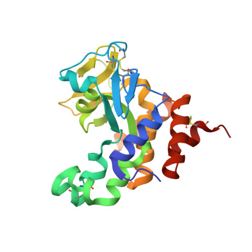
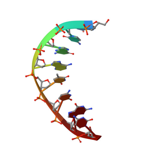
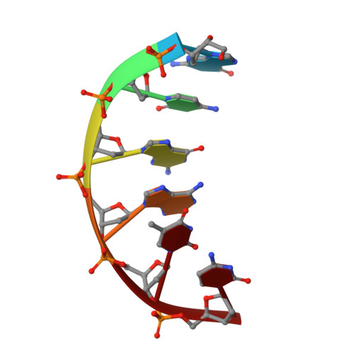
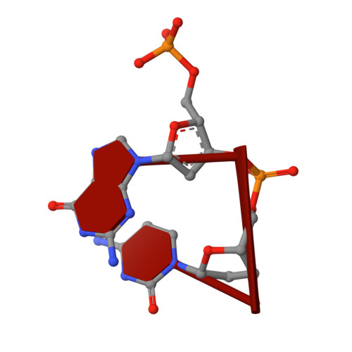
DNA binding and cleavage by the periplasmic nuclease Vvn: a novel structure with a known active site.

Li, C.-L.Hor, L.-I.Chang, Z.-F.Tsai, L.-C.Yang, W.-Z.Yuan, H.S.

(2003) EMBO J 22: 4014-4025

  • DOI: https://doi.org/10.1093/emboj/cdg377
  • Primary Citation Related Structures: 
    1OUO, 1OUP

  • PubMed Abstract: 

    The Vibrio vulnificus nuclease, Vvn, is a non-specific periplasmic nuclease capable of digesting DNA and RNA. The crystal structure of Vvn and that of Vvn mutant H80A in complex with DNA were resolved at 2.3 A resolution. Vvn has a novel mixed alpha/beta topology containing four disulfide bridges, suggesting that Vvn is not active under reducing conditions in the cytoplasm. The overall structure of Vvn shows no similarity to other endonucleases; however, a known 'betabetaalpha-metal' motif is identified in the central cleft region. The crystal structure of the mutant Vvn-DNA complex demonstrates that Vvn binds mainly at the minor groove of DNA, resulting in duplex bending towards the major groove by approximately 20 degrees. Only the DNA phosphate backbones make hydrogen bonds with Vvn, suggesting a structural basis for its sequence-independent recognition of DNA and RNA. Based on the enzyme-substrate and enzyme-product structures observed in the mutant Vvn-DNA crystals, a catalytic mechanism is proposed. This structural study suggests that Vvn hydrolyzes DNA by a general single-metal ion mechanism, and indicates how non-specific DNA-binding proteins may recognize DNA.


  • Organizational Affiliation
    • Institute of Biochemistry and Molecular Biology, College of Medicine, National Taiwan University, Institute of Molecular Biology, Academia Sinica, Taipei, Taiwan, Republic of China.

Macromolecule Content 

  • Total Structure Weight: 59.25 kDa 
  • Atom Count: 4,425 
  • Modeled Residue Count: 455 
  • Deposited Residue Count: 458 
  • Unique protein chains: 1
  • Unique nucleic acid chains: 3

Macromolecules


Find similar proteins by:|  3D Structure
Entity ID: 4
MoleculeChains  Sequence LengthOrganismDetailsImage
NucleaseF [auth A],
G [auth B]
213Vibrio vulnificusMutation(s): 1 
EC: 3.1
UniProt
Find proteins for Q7MHK3 (Vibrio vulnificus (strain YJ016))
Explore Q7MHK3 
Go to UniProtKB:  Q7MHK3
Entity Groups
Sequence Clusters30% Identity50% Identity70% Identity90% Identity95% Identity100% Identity
UniProt GroupQ7MHK3
Sequence Annotations
Expand
Reference Sequence
Find similar nucleic acids by:  Sequence
Entity ID: 1
MoleculeChains LengthOrganismImage
5'-D(*GP*CP*GP*AP*TP*CP*GP*C)-3'A [auth C],
B [auth D],
E [auth F]
8N/A
Sequence Annotations
Expand
Reference Sequence
Find similar nucleic acids by:  Sequence
Entity ID: 2
MoleculeChains LengthOrganismImage
5'-D(*GP*CP*GP*AP*TP*C)-3'C [auth E]6N/A
Sequence Annotations
Expand
Reference Sequence
Find similar nucleic acids by:  Sequence
Entity ID: 3
MoleculeChains LengthOrganismImage
5'-D(P*GP*C)-3'D [auth G]2N/A
Sequence Annotations
Expand
Reference Sequence

Experimental Data & Validation

Experimental Data

  • Method: X-RAY DIFFRACTION
  • Resolution: 2.30 Å
  • R-Value Free:  0.260 (Depositor), 0.260 (DCC) 
  • R-Value Work:  0.191 (Depositor), 0.191 (DCC) 
Space Group: P 1 21 1
Unit Cell:
Length ( Å )Angle ( ˚ )
a = 53.74α = 90
b = 50.11β = 97.53
c = 89.61γ = 90
Software Package:
Software NamePurpose
CNSrefinement
DENZOdata reduction
SCALEPACKdata scaling
CNSphasing

Structure Validation

View Full Validation Report


Entry History 

Deposition Data

Revision History  (Full details and data files)

  • Version 1.0: 2003-08-05
    Type: Initial release
  • Version 1.1: 2008-04-29
    Changes: Version format compliance
  • Version 1.2: 2011-07-13
    Changes: Version format compliance
  • Version 1.3: 2017-10-11
    Changes: Refinement description
  • Version 1.4: 2021-11-10
    Changes: Database references, Derived calculations
  • Version 1.5: 2024-04-03
    Changes: Data collection, Refinement description
  • Version 1.6: 2024-10-30
    Changes: Structure summary