1OT5

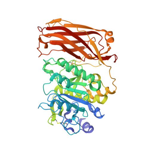
The 2.4 Angstrom Crystal Structure of Kex2 in complex with a peptidyl-boronic acid inhibitor


Experimental Data Snapshot

  • Method: X-RAY DIFFRACTION
  • Resolution: 2.40 Å
  • R-Value Free: 0.245 
  • R-Value Work: 0.209 
  • R-Value Observed: 0.218 

wwPDB Validation   3D Report Full Report


Ligand Structure Quality Assessment 


This is version 2.1 of the entry. See complete history


Literature

2.4 A Resolution Crystal Structure of the Prototypical Hormone-Processing Protease Kex2 in Complex with an Ala-Lys-Arg Boronic Acid Inhibitor

Holyoak, T.Wilson, M.A.Fenn, T.D.Kettner, C.A.Petsko, G.A.Fuller, R.S.Ringe, D.

(2003) Biochemistry 42: 6709-6718

  • DOI: https://doi.org/10.1021/bi034434t
  • Primary Citation of Related Structures:  
    1OT5

  • PubMed Abstract: 

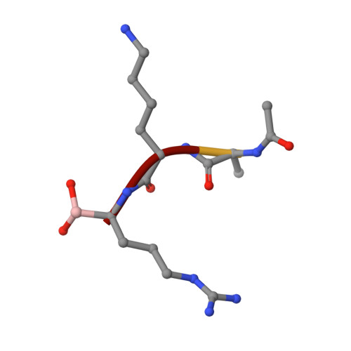
    This paper reports the first structure of a member of the Kex2/furin family of eukaryotic pro-protein processing proteases, which cleave sites consisting of pairs or clusters of basic residues. Reported is the 2.4 A resolution crystal structure of the two-domain protein ssKex2 in complex with an Ac-Ala-Lys-boroArg inhibitor (R = 20.9%, R(free) = 24.5%). The Kex2 proteolytic domain is similar in its global fold to the subtilisin-like superfamily of degradative proteases. Analysis of the complex provides a structural basis for the extreme selectivity of this enzyme family that has evolved from a nonspecific subtilisin-like ancestor. The P-domain of ssKex2 has a novel jelly roll like fold consisting of nine beta strands and may potentially be involved, along with the buried Ca(2+) ion, in creating the highly determined binding site for P(1) arginine.


  • Organizational Affiliation

    Rosenstiel Basic Medical Sciences Research Center, Brandeis University, Waltham, Massachusetts 02454, USA.


Macromolecules
Find similar proteins by:  (by identity cutoff)  |  3D Structure
Entity ID: 1
MoleculeChains Sequence LengthOrganismDetailsImage
Kexin
A, B
477Saccharomyces cerevisiaeMutation(s): 0 
Gene Names: KEX2 OR QDS1 OR YNL238W OR N1122
EC: 3.4.21.61
UniProt
Find proteins for P13134 (Saccharomyces cerevisiae (strain ATCC 204508 / S288c))
Explore P13134 
Go to UniProtKB:  P13134
Entity Groups  
Sequence Clusters30% Identity50% Identity70% Identity90% Identity95% Identity100% Identity
UniProt GroupP13134
Glycosylation
Glycosylation Sites: 2Go to GlyGen: P13134-1
Sequence Annotations
Expand
  • Reference Sequence

Find similar proteins by:  Sequence   |   3D Structure  

Entity ID: 2
MoleculeChains Sequence LengthOrganismDetailsImage
Ac-Ala-Lys-boroArg N-acetylated boronic acid peptide inhibitor
C, D
4N/AMutation(s): 0 
Sequence Annotations
Expand
  • Reference Sequence
Oligosaccharides

Help

Entity ID: 3
MoleculeChains Length2D Diagram Glycosylation3D Interactions
2-acetamido-2-deoxy-beta-D-glucopyranose-(1-4)-2-acetamido-2-deoxy-beta-D-glucopyranose
E, F
2N-Glycosylation
Glycosylation Resources
GlyTouCan:  G42666HT
GlyCosmos:  G42666HT
GlyGen:  G42666HT
Small Molecules
Ligands 2 Unique
IDChains Name / Formula / InChI Key2D Diagram3D Interactions
NAG
Query on NAG

Download Ideal Coordinates CCD File 
G [auth A],
K [auth B]
2-acetamido-2-deoxy-beta-D-glucopyranose
C8 H15 N O6
OVRNDRQMDRJTHS-FMDGEEDCSA-N
CA
Query on CA

Download Ideal Coordinates CCD File 
H [auth A]
I [auth A]
J [auth A]
L [auth B]
M [auth B]
H [auth A],
I [auth A],
J [auth A],
L [auth B],
M [auth B],
N [auth B]
CALCIUM ION
Ca
BHPQYMZQTOCNFJ-UHFFFAOYSA-N
Modified Residues  1 Unique
IDChains TypeFormula2D DiagramParent
BOR
Query on BOR
C, D
PEPTIDE-LIKEC5 H15 B N4 O2ARG
Experimental Data & Validation

Experimental Data

  • Method: X-RAY DIFFRACTION
  • Resolution: 2.40 Å
  • R-Value Free: 0.245 
  • R-Value Work: 0.209 
  • R-Value Observed: 0.218 
  • Space Group: P 65 2 2
Unit Cell:
Length ( Å )Angle ( ˚ )
a = 113.844α = 90
b = 113.844β = 90
c = 370.218γ = 120
Software Package:
Software NamePurpose
CNSrefinement
HKL-2000data reduction
SCALEPACKdata scaling
SOLVEphasing
RESOLVEphasing

Structure Validation

View Full Validation Report


Ligand Structure Quality Assessment 


Entry History 

Deposition Data

Revision History  (Full details and data files)

  • Version 1.0: 2003-06-17
    Type: Initial release
  • Version 1.1: 2008-04-29
    Changes: Version format compliance
  • Version 1.2: 2011-07-13
    Changes: Atomic model, Database references, Derived calculations, Non-polymer description, Structure summary, Version format compliance
  • Version 1.3: 2012-12-12
    Changes: Other
  • Version 1.4: 2013-02-20
    Changes: Other
  • Version 1.5: 2016-12-14
    Changes: Structure summary
  • Version 2.0: 2020-07-29
    Type: Remediation
    Reason: Carbohydrate remediation
    Changes: Atomic model, Data collection, Derived calculations, Structure summary
  • Version 2.1: 2024-10-30
    Changes: Data collection, Database references, Structure summary