1OMS | pdb_00001oms

Structure determination by MAD: E.coli Trigger Factor binding at the ribosomal exit tunnel.


Experimental Data Snapshot

  • Method: X-RAY DIFFRACTION
  • Resolution: 2.30 Å
  • R-Value Free: 
    0.238 (Depositor), 0.205 (DCC) 
  • R-Value Work: 
    0.208 (Depositor), 0.204 (DCC) 
  • R-Value Observed: 
    0.208 (Depositor) 

wwPDB Validation 3D Report Full Report

Validation slider image for 1OMS

This is version 2.0 of the entry. See complete history

Literature

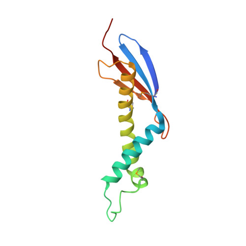
Chaperone binding at the ribosomal exit tunnel.

Kristensen, O.Gajhede, M.

(2003) Structure 11: 1547-1556

  • DOI: https://doi.org/10.1016/j.str.2003.11.003
  • Primary Citation Related Structures: 
    1OMS, 1P9Y

  • PubMed Abstract: 

    The exit tunnel region of the ribosome is well established as a focal point for interaction between the components that guide the fate of nascent polypeptides. One of these, the chaperone trigger factor (TF), associates with the 50S ribosomal subunit through its N-terminal domain. Targeting of TF to ribosomes is crucial to achieve its remarkable efficiency in protein folding. A similar tight coupling to translation is found in signal recognition particle (SRP)-dependent protein translocation. Here, we report crystal structures of the E. coli TF ribosome binding domain. TF is structurally related to the Hsp33 chaperone but has a prominent ribosome anchor located as a tip of the molecule. This tip includes the previously established unique TF signature motif. Comparison reveals that this feature is not found in SRP structures. We identify a conserved helical kink as a hallmark of the TF structure that is most likely critical to ensure ribosome association.


  • Organizational Affiliation
    • Structural Biology, Department of Medicinal Chemistry, The Danish University of Pharmaceutical Sciences, Universitetsparken 2, DK-2100 Copenhagen, Denmark. ok@dfh.dk

Macromolecule Content 

  • Total Structure Weight: 42.89 kDa 
  • Atom Count: 3,065 
  • Modeled Residue Count: 349 
  • Deposited Residue Count: 363 
  • Unique protein chains: 1

Macromolecules

Find similar proteins by:|  3D Structure
Entity ID: 1
MoleculeChains  Sequence LengthOrganismDetailsImage
Trigger Factor
A, B, C
121Escherichia coliMutation(s): 0 
Gene Names: tig
EC: 5.2.1.8
UniProt
Find proteins for P0A850 (Escherichia coli (strain K12))
Explore P0A850 
Go to UniProtKB:  P0A850
Entity Groups
Sequence Clusters30% Identity50% Identity70% Identity90% Identity95% Identity100% Identity
UniProt GroupP0A850
Sequence Annotations
Expand
Reference Sequence

Small Molecules

Ligands 4 Unique
IDChains Name / Formula / InChI Key2D Diagram3D Interactions
PG4

Query on PG4


Download:Ideal Coordinates CCD File
M [auth B],
N [auth B],
T [auth C],
U [auth C],
V [auth C]
TETRAETHYLENE GLYCOL
C8 H18 O5
UWHCKJMYHZGTIT-UHFFFAOYSA-N
SO4

Query on SO4


Download:Ideal Coordinates CCD File
D [auth A]
E [auth A]
F [auth A]
G [auth A]
H [auth B]
D [auth A],
E [auth A],
F [auth A],
G [auth A],
H [auth B],
I [auth B],
J [auth B],
K [auth B],
L [auth B],
P [auth C],
Q [auth C],
R [auth C],
S [auth C]
SULFATE ION
O4 S
QAOWNCQODCNURD-UHFFFAOYSA-L
GOL

Query on GOL


Download:Ideal Coordinates CCD File
W [auth C]GLYCEROL
C3 H8 O3
PEDCQBHIVMGVHV-UHFFFAOYSA-N
SO2

Query on SO2


Download:Ideal Coordinates CCD File
O [auth B]SULFUR DIOXIDE
O2 S
RAHZWNYVWXNFOC-UHFFFAOYSA-N
Modified Residues  1 Unique
IDChains TypeFormula2D DiagramParent
MSE
Query on MSE
A, B, C
L-PEPTIDE LINKINGC5 H11 N O2 SeMET

Experimental Data & Validation

Experimental Data

  • Method: X-RAY DIFFRACTION
  • Resolution: 2.30 Å
  • R-Value Free:  0.238 (Depositor), 0.205 (DCC) 
  • R-Value Work:  0.208 (Depositor), 0.204 (DCC) 
  • R-Value Observed: 0.208 (Depositor) 
Space Group: H 3 2
Unit Cell:
Length ( Å )Angle ( ˚ )
a = 71.644α = 90
b = 71.644β = 90
c = 375.409γ = 120
Software Package:
Software NamePurpose
CNSrefinement
MOSFLMdata reduction
CCP4data scaling
SHARPphasing

Structure Validation

View Full Validation Report


Entry History 

Deposition Data

Revision History  (Full details and data files)

  • Version 1.0: 2003-12-16
    Type: Initial release
  • Version 1.1: 2008-04-29
    Changes: Version format compliance
  • Version 1.2: 2011-07-13
    Changes: Non-polymer description, Version format compliance
  • Version 2.0: 2024-11-13
    Changes: Atomic model, Data collection, Database references, Derived calculations, Structure summary