1OF6 | pdb_00001of6

crystal structure of the tyrosine-regulated 3-deoxy-d-arabino-heptulosonate-7-phosphate synthase from saccharomyces cerevisiae complexed with tyrosine and manganese


Experimental Data Snapshot

  • Method: X-RAY DIFFRACTION
  • Resolution: 2.10 Å
  • R-Value Free: 
    0.237 (Depositor), 0.246 (DCC) 
  • R-Value Work: 
    0.195 (Depositor) 

Starting Model: experimental
View more details

wwPDB Validation 3D Report Full Report

Validation slider image for 1OF6

Ligand Structure Quality Assessment 


This is version 1.3 of the entry. See complete history

Literature

Crystal Structure of the Double Complex of the Tyrosine Sensitive Dahp Synthase from Yeast

Koenig, V.Pfeil, A.Heinrich, G.Braus, G.Schneider, T.R.

To be published.

Macromolecule Content 

  • Total Structure Weight: 320.27 kDa 
  • Atom Count: 21,605 
  • Modeled Residue Count: 2,770 
  • Deposited Residue Count: 2,960 
  • Unique protein chains: 1

Macromolecules

Find similar proteins by:|  3D Structure
Entity ID: 1
MoleculeChains  Sequence LengthOrganismDetailsImage
PHOSPHO-2-DEHYDRO-3-DEOXYHEPTONATE ALDOLASE, TYROSINE-INHIBITED
A, B, C, D, E
A, B, C, D, E, F, G, H
370Saccharomyces cerevisiaeMutation(s): 0 
EC: 4.1.2.15 (PDB Primary Data), 2.5.1.54 (UniProt)
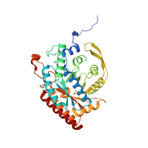
UniProt
Find proteins for P32449 (Saccharomyces cerevisiae (strain ATCC 204508 / S288c))
Explore P32449 
Go to UniProtKB:  P32449
Entity Groups
Sequence Clusters30% Identity50% Identity70% Identity90% Identity95% Identity100% Identity
UniProt GroupP32449
Sequence Annotations
Expand
Reference Sequence

Small Molecules

Ligands 2 Unique
IDChains Name / Formula / InChI Key2D Diagram3D Interactions
DTY

Query on DTY


Download:Ideal Coordinates CCD File
J [auth A]
L [auth B]
N [auth C]
P [auth D]
R [auth E]
J [auth A],
L [auth B],
N [auth C],
P [auth D],
R [auth E],
T [auth F],
V [auth G],
X [auth H]
D-TYROSINE
C9 H11 N O3
OUYCCCASQSFEME-MRVPVSSYSA-N
MN

Query on MN


Download:Ideal Coordinates CCD File
I [auth A]
K [auth B]
M [auth C]
O [auth D]
Q [auth E]
I [auth A],
K [auth B],
M [auth C],
O [auth D],
Q [auth E],
S [auth F],
U [auth G],
W [auth H]
MANGANESE (II) ION
Mn
WAEMQWOKJMHJLA-UHFFFAOYSA-N

Experimental Data & Validation

Experimental Data

  • Method: X-RAY DIFFRACTION
  • Resolution: 2.10 Å
  • R-Value Free:  0.237 (Depositor), 0.246 (DCC) 
  • R-Value Work:  0.195 (Depositor) 
Space Group: P 1
Unit Cell:
Length ( Å )Angle ( ˚ )
a = 82.129α = 64.71
b = 94.696β = 85.51
c = 104.843γ = 75.61
Software Package:
Software NamePurpose
REFMACrefinement
DENZOdata reduction
SCALEPACKdata scaling
EPMRphasing

Structure Validation

View Full Validation Report


Ligand Structure Quality Assessment 


Entry History 

Deposition Data

Revision History  (Full details and data files)

  • Version 1.0: 2004-05-06
    Type: Initial release
  • Version 1.1: 2014-02-05
    Changes: Database references, Derived calculations, Non-polymer description, Other, Source and taxonomy, Version format compliance
  • Version 1.2: 2019-07-24
    Changes: Data collection
  • Version 1.3: 2023-12-13
    Changes: Data collection, Database references, Derived calculations, Other, Refinement description