1OBM | pdb_00001obm

RECOMBINANT SPERM WHALE MYOGLOBIN 29F/64Q/68F/122N MUTANT (MET)


Experimental Data Snapshot

  • Method: X-RAY DIFFRACTION
  • Resolution: 1.85 Å
  • R-Value Work: 
    0.168 (Depositor) 
  • R-Value Observed: 
    0.168 (Depositor) 

wwPDB Validation 3D Report Full Report

Validation slider image for 1OBM

This is version 1.4 of the entry. See complete history

Literature

Solution and crystal structures of a sperm whale myoglobin triple mutant that mimics the sulfide-binding hemoglobin from Lucina pectinata.

Nguyen, B.D.Zhao, X.Vyas, K.La Mar, G.N.Lile, R.A.Brucker, E.A.Phillips Jr., G.N.Olson, J.S.Wittenberg, J.B.

(1998) J Biological Chem 273: 9517-9526

  • DOI: https://doi.org/10.1074/jbc.273.16.9517
  • Primary Citation Related Structures: 
    1OBM

  • PubMed Abstract: 

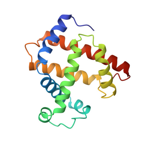
    The bivalve mollusc Lucina pectinata harbors sulfide-oxidizing chemoautotrophic bacteria and expresses a monomeric hemoglobin I, HbI, with normal O2, but extraordinarily high sulfide affinity. The crystal structure of aquomet Lucina HbI has revealed an active site with three residues not commonly found in vertebrate globins: Phe(B10), Gln(E7), and Phe(E11) (Rizzi, M., Wittenberg, J. B., Coda, A., Fasano, M., Ascenzi, P., and Bolognesi, M. (1994) J. Mol. Biol. 244, 86-89). Engineering these three residues into sperm whale myoglobin results in a triple mutant with approximately 700-fold higher sulfide affinity than for wild-type. The single crystal x-ray structure of the aquomet derivative of the myoglobin triple mutant and the solution 1H NMR active site structures of the cyanomet derivatives of both the myoglobin mutant and Lucina HbI have been determined to examine further the structural origin of their unusually high sulfide affinities. The major differences in the distal pocket is that in the aquomet form the carbonyl of Gln64(E7) serves as a H-bond acceptor, whereas in the cyanomet form the amido group acts as H-bond donor to the bound ligand. Phe68(E11) is rotated approximately 90 degrees about chi2 and located approximately 1-2 A closer to the iron atom in the myoglobin triple mutant relative to its conformation in Lucina HbI. The change in orientation potentially eliminates the stabilizing interaction with sulfide and, together with the decrease in size of the distal pocket, accounts for the 7-fold lower sulfide affinity of the myoglobin mutant compared with that of Lucina HbI.


  • Organizational Affiliation
    • Department of Chemistry, University of California, Davis, California 95616, USA.

Macromolecule Content 

  • Total Structure Weight: 18.15 kDa 
  • Atom Count: 1,431 
  • Modeled Residue Count: 154 
  • Deposited Residue Count: 154 
  • Unique protein chains: 1

Macromolecules

Find similar proteins by:|  3D Structure
Entity ID: 1
MoleculeChains  Sequence LengthOrganismDetailsImage
MYOGLOBIN154Physeter macrocephalusMutation(s): 4 
EC: 1.11.1 (UniProt), 1.7 (UniProt)
UniProt
Find proteins for P02185 (Physeter macrocephalus)
Explore P02185 
Go to UniProtKB:  P02185
Entity Groups
Sequence Clusters30% Identity50% Identity70% Identity90% Identity95% Identity100% Identity
UniProt GroupP02185
Sequence Annotations
Expand
Reference Sequence

Small Molecules

Ligands 2 Unique
IDChains Name / Formula / InChI Key2D Diagram3D Interactions
HEM

Query on HEM


Download:Ideal Coordinates CCD File
C [auth A]PROTOPORPHYRIN IX CONTAINING FE
C34 H32 Fe N4 O4
KABFMIBPWCXCRK-RGGAHWMASA-L
SO4

Query on SO4


Download:Ideal Coordinates CCD File
B [auth A]SULFATE ION
O4 S
QAOWNCQODCNURD-UHFFFAOYSA-L

Experimental Data & Validation

Experimental Data

  • Method: X-RAY DIFFRACTION
  • Resolution: 1.85 Å
  • R-Value Work:  0.168 (Depositor) 
  • R-Value Observed: 0.168 (Depositor) 
Space Group: P 6
Unit Cell:
Length ( Å )Angle ( ˚ )
a = 91.39α = 90
b = 91.39β = 90
c = 45.75γ = 120
Software Package:
Software NamePurpose
X-PLORmodel building
X-PLORrefinement
XDSdata reduction
XDSdata scaling
X-PLORphasing

Structure Validation

View Full Validation Report


Entry History 

Deposition Data

Revision History  (Full details and data files)

  • Version 1.0: 1998-04-08
    Type: Initial release
  • Version 1.1: 2008-03-24
    Changes: Version format compliance
  • Version 1.2: 2011-07-13
    Changes: Version format compliance
  • Version 1.3: 2021-11-03
    Changes: Database references, Derived calculations, Other
  • Version 1.4: 2024-02-14
    Changes: Data collection