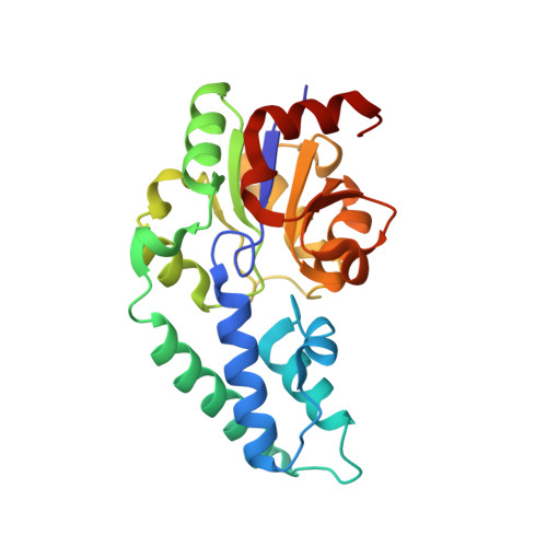
1O08

Structure of Pentavalent Phosphorous Intermediate of an Enzyme Catalyzed Phosphoryl transfer Reaction observed on cocrystallization with Glucose 1-phosphate


Experimental Data Snapshot

  • Method: X-RAY DIFFRACTION
  • Resolution: 1.20 Å
  • R-Value Free: 0.179 
  • R-Value Work: 0.141 
  • R-Value Observed: 0.141 

Starting Model: experimental
View more details

wwPDB Validation   3D Report Full Report


Ligand Structure Quality Assessment 


This is version 2.1 of the entry. See complete history


Literature

The pentacovalent phosphorus intermediate of a phosphoryl transfer reaction.

Lahiri, S.D.Zhang, G.Dunaway-Mariano, D.Allen, K.N.

(2003) Science 299: 2067-2071

  • DOI: https://doi.org/10.1126/science.1082710
  • Primary Citation of Related Structures:  
    1O03, 1O08

  • PubMed Abstract: 

    Enzymes provide enormous rate enhancements, unmatched by any other type of catalyst. The stabilization of high-energy states along the reaction coordinate is the crux of the catalytic power of enzymes. We report the atomic-resolution structure of a high-energy reaction intermediate stabilized in the active site of an enzyme. Crystallization of phosphorylated beta-phosphoglucomutase in the presence of the Mg(II) cofactor and either of the substrates glucose 1-phosphate or glucose 6-phosphate produced crystals of the enzyme-Mg(II)-glucose 1,6-(bis)phosphate complex, which diffracted x-rays to 1.2 and 1.4 angstroms, respectively. The structure reveals a stabilized pentacovalent phosphorane formed in the phosphoryl transfer from the C(1)O of glucose 1,6-(bis)phosphate to the nucleophilic Asp8 carboxylate.


  • Organizational Affiliation

    Department of Physiology and Biophysics, Boston University School of Medicine, Boston, MA 02118-2394, USA.


Macromolecules
Find similar proteins by:  (by identity cutoff)  |  3D Structure
Entity ID: 1
MoleculeChains Sequence LengthOrganismDetailsImage
beta-phosphoglucomutase221Lactococcus lactisMutation(s): 0 
EC: 5.4.2.6
UniProt
Find proteins for P71447 (Lactococcus lactis subsp. lactis (strain IL1403))
Explore P71447 
Go to UniProtKB:  P71447
Entity Groups  
Sequence Clusters30% Identity50% Identity70% Identity90% Identity95% Identity100% Identity
UniProt GroupP71447
Sequence Annotations
Expand
  • Reference Sequence
Small Molecules
Ligands 2 Unique
IDChains Name / Formula / InChI Key2D Diagram3D Interactions
G16
Query on G16

Download Ideal Coordinates CCD File 
B [auth A]1,6-di-O-phosphono-alpha-D-glucopyranose
C6 H13 O12 P2
RWHOZGRAXYWRNX-VFUOTHLCSA-M
MG
Query on MG

Download Ideal Coordinates CCD File 
C [auth A]MAGNESIUM ION
Mg
JLVVSXFLKOJNIY-UHFFFAOYSA-N
Experimental Data & Validation

Experimental Data

  • Method: X-RAY DIFFRACTION
  • Resolution: 1.20 Å
  • R-Value Free: 0.179 
  • R-Value Work: 0.141 
  • R-Value Observed: 0.141 
  • Space Group: P 21 21 21
Unit Cell:
Length ( Å )Angle ( ˚ )
a = 36.939α = 90
b = 54.297β = 90
c = 104.68γ = 90
Software Package:
Software NamePurpose
SHELXmodel building
SHELXL-97refinement
DENZOdata reduction
SCALEPACKdata scaling
SHELXphasing

Structure Validation

View Full Validation Report


Ligand Structure Quality Assessment 


Entry History 

Deposition Data

Revision History  (Full details and data files)

  • Version 1.0: 2003-03-18
    Type: Initial release
  • Version 1.1: 2008-04-29
    Changes: Version format compliance
  • Version 1.2: 2011-07-13
    Changes: Version format compliance
  • Version 1.3: 2020-07-29
    Type: Remediation
    Reason: Carbohydrate remediation
    Changes: Advisory, Data collection, Derived calculations, Structure summary
  • Version 2.0: 2024-04-03
    Changes: Atomic model, Data collection, Database references, Derived calculations, Refinement description, Structure summary
  • Version 2.1: 2024-10-16
    Changes: Structure summary