1NVI | pdb_00001nvi

Orthorhombic Crystal Form of Molybdopterin Synthase


Experimental Data Snapshot

  • Method: X-RAY DIFFRACTION
  • Resolution: 1.90 Å
  • R-Value Free: 
    0.206 (Depositor), 0.212 (DCC) 
  • R-Value Work: 
    0.175 (Depositor), 0.187 (DCC) 
  • R-Value Observed: 
    0.177 (Depositor) 

Starting Model: experimental
View more details

wwPDB Validation 3D Report Full Report

Validation slider image for 1NVI

This is version 1.4 of the entry. See complete history

Literature

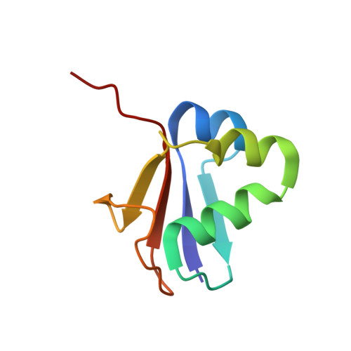
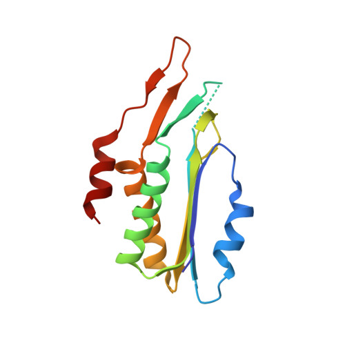
Structural Studies of Molybdopterin Synthase Provide Insights into its Catalytic Mechanism

Rudolph, M.J.Wuebbens, M.M.Turque, O.Rajagopalan, K.V.Schindelin, H.

(2003) J Biological Chem 278: 14514-14522

  • DOI: https://doi.org/10.1074/jbc.M300449200
  • Primary Citation Related Structures: 
    1NVI, 1NVJ

  • PubMed Abstract: 

    Molybdenum cofactor biosynthesis is an evolutionarily conserved pathway present in eubacteria, archaea, and eukaryotes, including humans. Genetic deficiencies of enzymes involved in cofactor biosynthesis in humans lead to a severe and usually fatal disease. The molybdenum cofactor contains a tricyclic pyranopterin, termed molybdopterin, that bears the cis-dithiolene group responsible for molybdenum ligation. The dithiolene group of molybdopterin is generated by molybdopterin synthase, which consists of a large (MoaE) and small (MoaD) subunit. The crystal structure of molybdopterin synthase revealed a heterotetrameric enzyme in which the C terminus of each MoaD subunit is deeply inserted into a MoaE subunit to form the active site. In the activated form of the enzyme, the MoaD C terminus is present as a thiocarboxylate. The present study identified the position of the thiocarboxylate sulfur by exploiting the anomalous signal originating from the sulfur atom. The structure of molybdopterin synthase in a novel crystal form revealed a binding pocket for the terminal phosphate of molybdopterin, the product of the enzyme, and suggested a binding site for the pterin moiety present in precursor Z and molybdopterin. Finally, the crystal structure of the MoaE homodimer provides insights into the conformational changes accompanying binding of the MoaD subunit.


  • Organizational Affiliation
    • Department of Biochemistry and Cell Biology, State University of New York at Stony Brook, 11794-5215, USA.

Macromolecule Content 

  • Total Structure Weight: 26.23 kDa 
  • Atom Count: 1,989 
  • Modeled Residue Count: 223 
  • Deposited Residue Count: 231 
  • Unique protein chains: 2

Macromolecules

Find similar proteins by:|  3D Structure
Entity ID: 1
MoleculeChains  Sequence LengthOrganismDetailsImage
Molybdopterin converting factor subunit 1A [auth D]81Escherichia coliMutation(s): 1 
Gene Names: MOAD OR CHLA4 OR CHLM OR B0784
UniProt
Find proteins for P30748 (Escherichia coli (strain K12))
Explore P30748 
Go to UniProtKB:  P30748
Entity Groups
Sequence Clusters30% Identity50% Identity70% Identity90% Identity95% Identity100% Identity
UniProt GroupP30748
Sequence Annotations
Expand
Reference Sequence
Find similar proteins by:|  3D Structure
Entity ID: 2
MoleculeChains  Sequence LengthOrganismDetailsImage
Molybdopterin converting factor subunit 2B [auth E]150Escherichia coliMutation(s): 0 
Gene Names: MOAE OR CHLA5 OR B0785
EC: 2.8.1.12
UniProt
Find proteins for P30749 (Escherichia coli (strain K12))
Explore P30749 
Go to UniProtKB:  P30749
Entity Groups
Sequence Clusters30% Identity50% Identity70% Identity90% Identity95% Identity100% Identity
UniProt GroupP30749
Sequence Annotations
Expand
Reference Sequence

Experimental Data & Validation

Experimental Data

  • Method: X-RAY DIFFRACTION
  • Resolution: 1.90 Å
  • R-Value Free:  0.206 (Depositor), 0.212 (DCC) 
  • R-Value Work:  0.175 (Depositor), 0.187 (DCC) 
  • R-Value Observed: 0.177 (Depositor) 
Space Group: I 21 21 21
Unit Cell:
Length ( Å )Angle ( ˚ )
a = 44.578α = 90
b = 94.86β = 90
c = 137.44γ = 90
Software Package:
Software NamePurpose
REFMACrefinement
DENZOdata reduction
SCALEPACKdata scaling
AMoREphasing

Structure Validation

View Full Validation Report


Entry History 

Deposition Data

Revision History  (Full details and data files)

  • Version 1.0: 2003-05-06
    Type: Initial release
  • Version 1.1: 2008-04-29
    Changes: Version format compliance
  • Version 1.2: 2011-07-13
    Changes: Derived calculations, Version format compliance
  • Version 1.3: 2021-10-27
    Changes: Database references, Derived calculations
  • Version 1.4: 2023-08-16
    Changes: Data collection, Refinement description