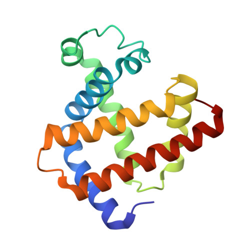
Crystal structures of ferrous horse heart myoglobin complexed with nitric oxide and nitrosoethane.
Copeland, D.M., West, A.H., Richter-Addo, G.B.(2003) Proteins 53: 182-192
- PubMed: 14517970 Search on PubMed
- DOI: https://doi.org/10.1002/prot.10495
- Primary Citation Related Structures:
1NPF, 1NPG - PubMed Abstract:
The interactions of nitric oxide (NO) and organic nitroso compounds with heme proteins are biologically important, and adduct formation between NO-containing compounds and myoglobin (Mb) have served as prototypical systems for studies of these interactions. We have prepared crystals of horse heart (hh) MbNO from nitrosylation of aqua-metMb crystals, and we have determined the crystal structure of hh MbNO at a resolution of 1.9 A. The Fe-N-O angle of 147 degrees in hh MbNO is larger than the corresponding 112 degrees angle previously determined from the crystal structure of sperm whale MbNO (Brucker et al., Proteins 1998;30:352-356) but is similar to the 150 degrees angle determined from a MS XAFS study of a frozen solution of hh MbNO (Rich et al., J Am Chem Soc 1998;120:10827-10836). The Fe-N(O) bond length of 2.0 A (this work) is longer than the 1.75 A distance determined from the XAFS study and suggests distal pocket influences on FeNO geometry. The nitrosyl N atom is located 3.0 A from the imidazole N(epsilon) atom of the distal His64 residue, suggesting electrostatic stabilization of the FeNO moiety by His64. The crystal structure of the nitrosoethane adduct of ferrous hh Mb was determined at a resolution of 1.7 A. The nitroso O atom of the EtNO ligand is located 2.7 A from the imidazole N(epsilon) atom of His64, suggesting a hydrogen bond interaction between these groups. To the best of our knowledge, the crystal structure of hh Mb(EtNO) is the first such determination of a nitrosoalkane adduct of a heme protein.
- Department of Chemistry and Biochemistry, University of Oklahoma, Norman, Oklahoma 73019, USA.
Organizational Affiliation: