1NNS | pdb_00001nns

L-asparaginase of E. coli in C2 space group and 1.95 A resolution


Experimental Data Snapshot

  • Method: X-RAY DIFFRACTION
  • Resolution: 1.95 Å
  • R-Value Free: 
    0.171 (Depositor), 0.173 (DCC) 
  • R-Value Work: 
    0.130 (Depositor), 0.132 (DCC) 
  • R-Value Observed: 
    0.132 (Depositor) 

Starting Model: experimental
View more details

wwPDB Validation 3D Report Full Report

Validation slider image for 1NNS

This is version 1.6 of the entry. See complete history

Literature

Structural comparison of Escherichia coli L-asparaginase in two monoclinic space groups.

Sanches, M.Barbosa, J.A.R.G.de Oliveira, R.T.Abrahao Neto, J.Polikarpov, I.

(2003) Acta Crystallogr D Biol Crystallogr 59: 416-422

  • DOI: https://doi.org/10.1107/s0907444902021200
  • Primary Citation Related Structures: 
    1NNS

  • PubMed Abstract: 

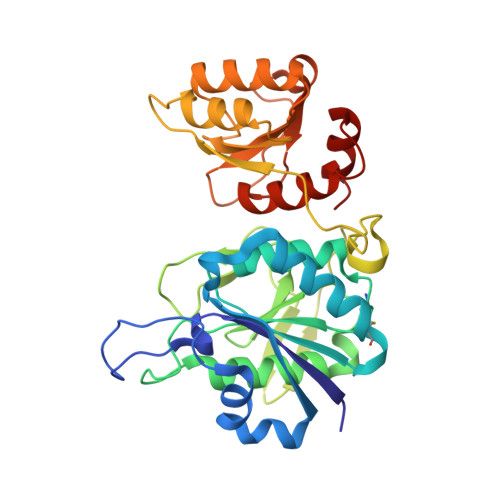
    The functional L-asparaginase from Escherichia coli is a homotetramer with a molecular weight of about 142 kDa. The X-ray structure of the enzyme, crystallized in a new form (space group C2) and refined to 1.95 A resolution, is compared with that of the previously determined crystal form (space group P2(1)). The asymmetric unit of the new crystal form contains an L-asparaginase dimer instead of the tetramer found in the previous crystal form. It is found that crystal contacts practically do not affect the conformation of the protein. It is shown that subunit C of the tetrameric form is in a conformation which is systematically different from that of all other subunits in both crystal forms. Major conformational differences are confined to the lid loop (residues 14-27). In addition, the stability of this globular protein is analyzed in terms of the interactions between hydrophobic parts of the subunits.


  • Organizational Affiliation
    • Grupo de Cristalografia, Departamento de Física em Sã;o Carlos, USP, Av. Trabalhador SãoCarlense 400, CEP 13560-970, São Carlos/SP, Brazil.

Macromolecule Content 

  • Total Structure Weight: 69.52 kDa 
  • Atom Count: 5,209 
  • Modeled Residue Count: 652 
  • Deposited Residue Count: 652 
  • Unique protein chains: 1

Macromolecules

Find similar proteins by:|  3D Structure
Entity ID: 1
MoleculeChains  Sequence LengthOrganismDetailsImage
L-asparaginase II
A, B
326Escherichia coliMutation(s): 0 
EC: 3.5.1.1
UniProt
Find proteins for P00805 (Escherichia coli (strain K12))
Explore P00805 
Go to UniProtKB:  P00805
Entity Groups
Sequence Clusters30% Identity50% Identity70% Identity90% Identity95% Identity100% Identity
UniProt GroupP00805
Sequence Annotations
Expand
Reference Sequence

Experimental Data & Validation

Experimental Data

  • Method: X-RAY DIFFRACTION
  • Resolution: 1.95 Å
  • R-Value Free:  0.171 (Depositor), 0.173 (DCC) 
  • R-Value Work:  0.130 (Depositor), 0.132 (DCC) 
  • R-Value Observed: 0.132 (Depositor) 
Space Group: C 1 2 1
Unit Cell:
Length ( Å )Angle ( ˚ )
a = 76.296α = 90
b = 134.617β = 110.51
c = 64.867γ = 90
Software Package:
Software NamePurpose
MAR345data collection
SCALEPACKdata scaling
AMoREphasing
REFMACrefinement

Structure Validation

View Full Validation Report


Entry History 

Deposition Data

Revision History  (Full details and data files)

  • Version 1.0: 2003-03-11
    Type: Initial release
  • Version 1.1: 2008-04-29
    Changes: Version format compliance
  • Version 1.2: 2011-07-13
    Changes: Advisory, Derived calculations, Version format compliance
  • Version 1.3: 2017-07-05
    Changes: Database references
  • Version 1.4: 2017-10-11
    Changes: Refinement description
  • Version 1.5: 2023-08-16
    Changes: Data collection, Database references, Derived calculations, Refinement description
  • Version 1.6: 2024-10-30
    Changes: Structure summary